Donor Prion Disease Precautions

Principle:

Prion diseases, including variant Creutzfeld-Jakob disease (mad cow disease), can be efficiently transmitted by transfusion of blood components.  Since there is a variable, often long (measured in months to years) incubation time before the disease is manifest, it is important to note individuals at high-risk and exclude them from the donor pool.

This is the August 2020 revision of the previous CBER recommendation on this topic.

Policy:

The following categories of individuals are permanently prohibited from donation (whole blood, apheresis, or organ):

  1. Anyone who received human growth hormone derived from human pituitary glands
  2. Anyone who received dura mater grafts
  3. Anyone from families with a history of Creutzfeld-Jakob Disease or other prion diseases (including variant Creutzfeld-Jakob—Mad Cow Disease, Gerstmann-Straussler-Scheinker Syndrome, Fatal Familial Insomnia, Kuru)
  4. Anyone with a family history of dementia or any other chronic neurologic illnesses
  5. Anyone with first-degree relatives (parent, sibling, children) who have a prion disease
  6. Anyone who has used (injected) bovine insulin of UK origin since 1980
  7. Defer indefinitely a donor who has spent five years or more cumulatively in France or Ireland from 1980 to 2001.
  8. Anyone having resided in the United Kingdom or visited there for a total of at least three months between 1980-96
  9. Anyone who received a blood component transfusion in the United Kingdom, France, or Ireland since 1980.

Definitions:

  1. For the purposes of this policy, the United Kingdom UK is defined as any one of the following areas:  England, Northern Ireland, Scotland, Wales, Isle of Man, Gibraltar, Channel Islands, or the Falkland Islands.
  2. France refers only to mainland France and NOT to French overseas departments, e.g. Martinique, French Guiana, Guadeloupe, Mayotte, and Réunion

All donors (except autologous) must be asked questions to rule out any of the above possibilities.

References:

  1. Recommendations to Reduce the Possible Risk of Transmission of Creutzfeld-Jakob Disease and Variant Creutzfeld-Jakob Disease by Blood and Blood Components, Guidance for Industry, US Department for Health and Human Services. FDA, Center for Biologics Evaluation and Research CBER, August, 2020
  2. Association Bulletin #02-2, American Association of Blood Banks, Bethesda, Maryland, USA, 8/3/02
  3. FDA Guidance for Industry:  Revised Preventive Measures to Reduce the Possible Risk of Transmission of Creutzfeld-Jakob and Variant Creutzfeld-Jakob Disease by Blood and Blood Products, FDA Guidance for Industry, 9/1/2002
  4. Belay E.D. and Schonberger L.B., The Public Health Impact of Prion Diseases, Annu. Rev. Public Health 2005. 26:191–212, Division of Viral and Rickettsial Diseases, Centers for Disease Control and Prevention, Atlanta Georgia,
  5. Standards for Blood Banks and Transfusion Services, Current Edition, AABB, Bethesda, MD, USA
  6. Guidelines to the Preparation, Use, and Quality Assurance of Blood Components, European Committee (Partial Agreement) on Blood Transfusion (CD-P-TS), Current Edition
Transfusion Reaction Workup Form

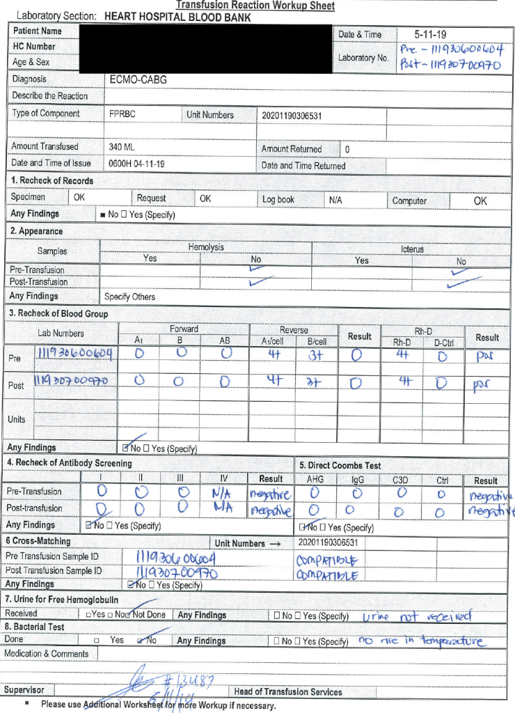
It is easy to regurgitate data, but the key to using it successfully is to organize to maximize pattern recognition.  Once a transfusion reaction has been called, the transfusion blood bank staff must expeditiously (STAT) perform testing to rule out acute hemolysis—one of the four fatal transfusion reactions (others being anaphylaxis, septic shock, TRALI/TACO).

The attached form was designed by my staff and me based on my experiences at several institutions.  It organizes the workup and helps ensure that all the testing and checks are performed.  The clinical area calling the reaction will provide a transfusion form that includes the vital signs (pre-, post-, and during the transfusion) as well as the symptoms.

With these forms, life-threatening hemolysis can be ruled out and further studies made to rule out other serious adverse reactions.

The data on this worksheet are entered into the Medinfo Hematos IIG Patient Module. The transfusion physician reviewing the workup will enter the type of reaction and recommendations. This will document the transfusion reaction for accreditation purposes.

10/10/20

Processes and Software Building 51: Donor D Typing

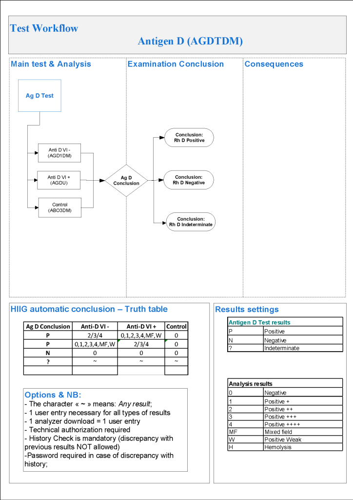
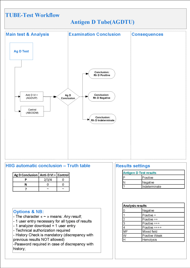
For donor D typing, I always had both automated and manual methods set up on the blood bank computer system Medinfo Hematos IIG.  The automated method had a bidirectional interface between Medinfo and the instrument.   Medinfo did not need a separate middleware.  A truth table was prepared for acceptable results for automatic interpretation.  Other results had to be manually interpreted by someone with the appropriate security level.

For donors, I wanted reagents sensitive for partial or mosaic D types since any D epitopes are potentially immunogenic.  With some reagents, this meant using a DVI+ reagent and others both DVI+ and DVI- reagents.

The manual testing option is structured similarly.  Within Medinfo, it is easy to change the methodology if the system is so built.  Thus, if the analyzer for D typing is down, the staff can select the manual methodology.  Likewise, if one testing center goes off-line, the world can be completed at another site—no need to repeat testing already completed from the first site.  This flexibility can apply to any test in system.

The manufacturer’s recommendations for the particular reagents in use were strictly followed.  One used the range {0, 1, 2, 3, 4, Mixed Field, Weak} as acceptable.  Another used {0, 2, 3, 4}.  Controls were included.  Most importantly, Medinfo can be configured for any set of reagent values.

The attached flows show two different testing systems as examples of what can be built in the Medinfo system.

9/10/20

Opinion: Continue Manual Data Collection During Therapeutic Apheresis Procedures

While I was  Division Head, Laboratory Information Systems LIS at my previous position, I was asked to use the hospital information system HIS to collect information during the procedure analogous to what was done for dialysis.

I thought of the logistics:  one apheresis nurse, one Spectra Optia machine, and one metal cage containing a theft-proof computer on a stand.  There was no room for the patient’s bed with all this equipment—the nurse could not move around comfortably.

Second, what I was presented was a hodge-podge of screens on the HIS that the apheresis had to maneuver back and forth between for each measurement—none of the data entry was on one screen!  Honestly, there wasn’t enough time to enter all the data between the screens AND look at the patient.

I remind everyone that therapeutic apheresis is not a benign procedure.  The patient may be critically ill.  The apheresis nurse must concentrate on the patient.  The HIS team was more interested in the data collection, even at the expense of the patient.

LIS had not been engaged in building the pathway and the HIS wanted us to follow the dialysis template.  They did not know that there are many types of therapeutic procedures, often with different data collection.  There is no one-size-fits-all screen!

I refused.  The nurse must concentrate on the patient, not the LCD screen.  To use the HIS would have been harmful to patient care in this situation.  We retained the manual, cellulose interface.  We scanned the manual data form and uploaded it into HIS.

Lessons to be learned:

  1. HIS must engage LIS, and in particular Transfusion Medicine, when building anything for the blood bank.  This is in accordance with international  accreditation standards.
  2. We must never lose sight that we are treating the patient, not the computer screen.  Especially in therapeutic apheresis, we must use the apheresis specialist nurse to monitor the clinical status of the patient, first and foremost!
  3. If the proposed computer process is worse than the manual process, keep the latter.

8/10/20

Processes and Software Building 50: Donor ABO Confirmatory Typing

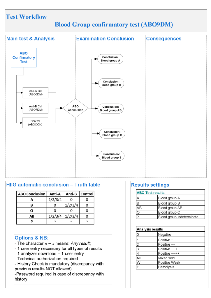
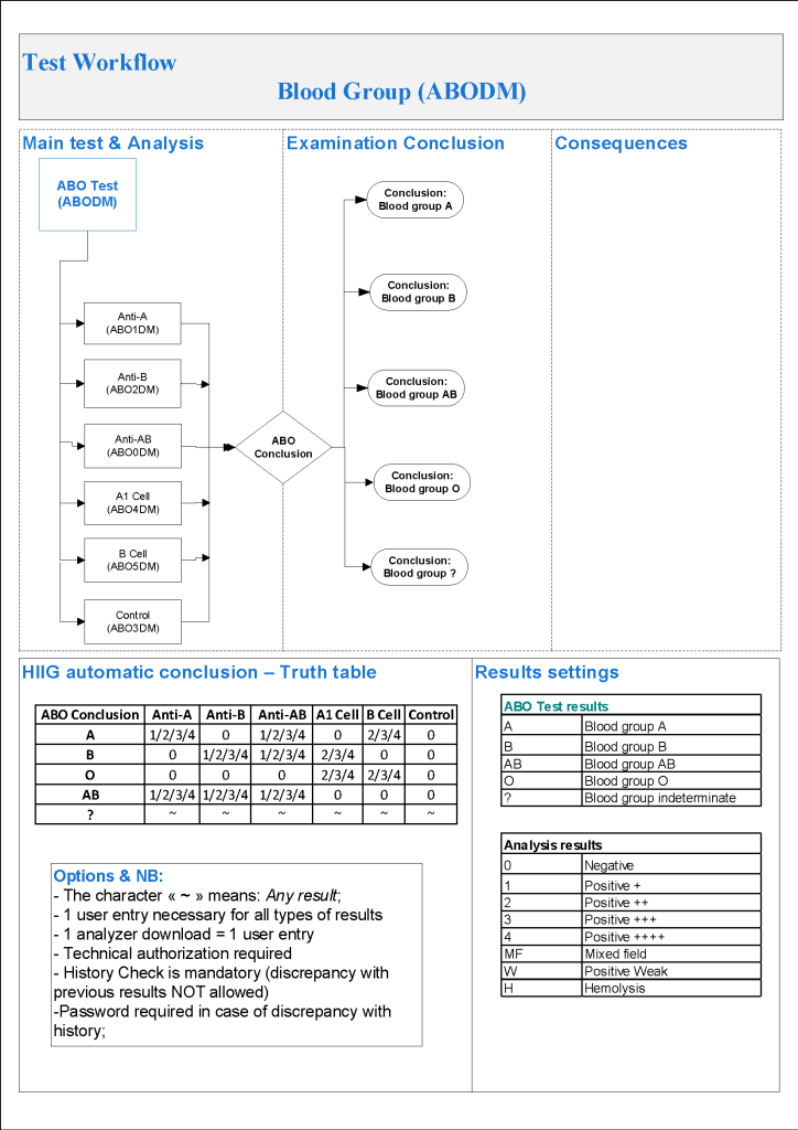
For donor ABO confirmatory typing, I always had both automated and manual methods set up on the blood bank computer system Medinfo Hematos IIG.  The automated method had a bidirectional interface between Medinfo and the instrument.   Medinfo did not need a separate middleware.  A truth table was prepared for acceptable results for automatic interpretation.  Other results had to be manually interpreted by someone with the appropriate security level.

The manual testing option is structured similarly.  Within Medinfo, it is easy to change the methodology if the system is so built.  Thus, if the analyzer for ABO typing is down, the staff can select the manual methodology.  Likewise, if one testing center goes off-line, the world can be completed at another site—no need to repeat testing already completed from the first site.  This flexibility can apply to any test in system.

The manufacturer’s recommendations for the particular reagents in use were strictly followed.  One used the range {0, 1, 2, 3, 4} as acceptable.  Another used {0, 2, 3, 4}.  Controls were included.  Most importantly, Medinfo can be configured for any set of reagent values.  Refer to the following flow diagram.

Confirmatory testing also includes D typing.  That will be considered in a future post.

6/10/20

Contacting the Transfusion Physician with Transfusion Reaction Workup Results

Principle:

The physician on-call for the blood bank requires a certain minimum amount of data to determine the significance of a suspected transfusion reaction and to decide if further testing is required.

Policy:

  1. DO NOT call the transfusion physician about a transfusion reaction until the following data is ready:
    1. Patient name and hospital number
    2. Patient age and diagnosis and location
    3. Previous transfusion history including antibodies and previous transfusion reactions
    4. Vital signs (BP, temperature, pulse, and respiratory rate) before AND after the transfusion
    5. Clinical symptoms (e.g. fever, chills, rash, urticaria, dyspnea, hematuria, etc.)
    6. Repeat ABO and D type on the post-transfusion specimen
    7. Direct antiglobulin/Coombs test (DAT) on the post-transfusion specimen
    8. DAT on the pretransfusion specimen if the post-transfusion specimen is DAT-positive
    9. Results of hemolysis check on pre- and post-transfusion plasma/serum
  2. The transfusion physician may order additional testing based on the above results.

My Opinion: Use Gamma-Heavy-Chain-Specific AHG Reagent

There are many types of antiglobulin reagent available which have differing specificities to immunoglobulins (e.g. whole molecule IgG, IgG-heavy-chain specific, IgM-heavy-chain-specific, IgA-heavy-chain-specific) and complement fragments (e.g. C3b, C3c, C3d).

For the purpose of antibody identification using the indirect antiglobulin test IAT, normally polyspecific, whole molecule IgG, and/or gamma-heavy-chain-specific reagents are used.

The purpose of antibody identification is normally to detect clinically significant antibodies—NOT ALL ANTIBODIES.  In a busy hospital blood bank, I am not routinely interested in detecting cold antibodies that do not react at 37C.

Polyspecific reagents will detect both complement and/or immunoglobulin and are commonly chosen.  However, the complement may detect insignificant cold antibodies that may obscure clinically significant IgG antibodies.

A whole-molecule IgG reagent is not really monospecific since it detects both heavy and light chains.  The light chains (kappa and lambda) are shared by all classes of immunoglobulin.  Thus, an IgM antibody (usually cold) may show weak reactivity—nonspecific cold antibodies may be detected!

My usual choice is to routinely use a gamma-heavy-chain-specific reagent.  I have been using this for many years and it expedites the workflow.  The likelihood of missing a clinically significant antibody is rare.  In my long career, I have only detected a small number of Kidd antibodies that required a polyspecific reagent with complement. 

If there is a nonspecific antibody, I will check the Jka and Jkb typings.  If either is negative, I will check for an antibody showing dosage and consider using a polyspecific reagent.  I have previously reported such an antibody in an earlier post—it is extremely rare!

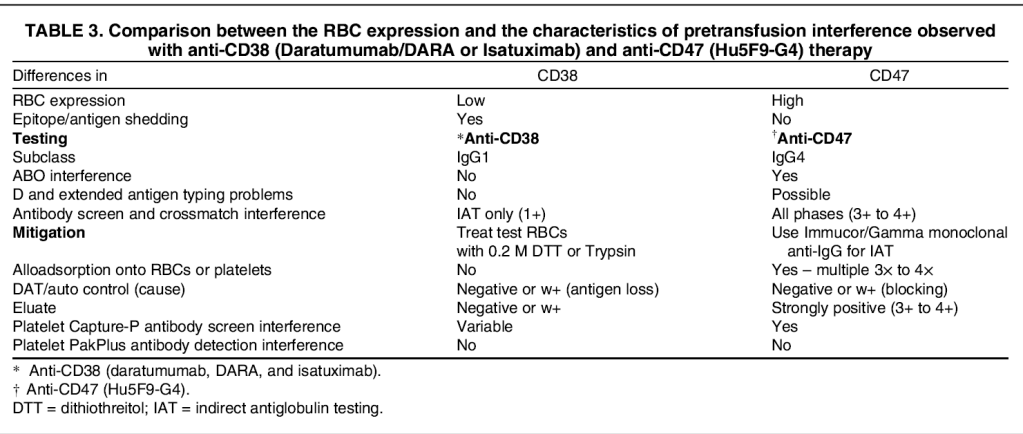
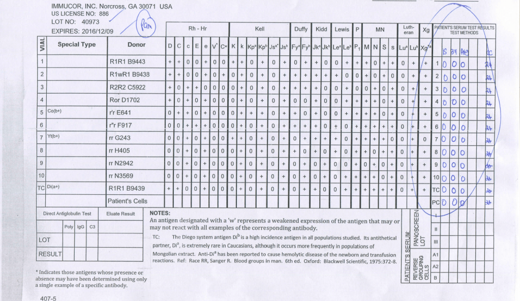
Another consideration is the detection of interference from a new chemotherapy reagent, anti-CD47.  This nonspecific reactivity will be eliminated by using a gamma-heavy-chain specific reagent:

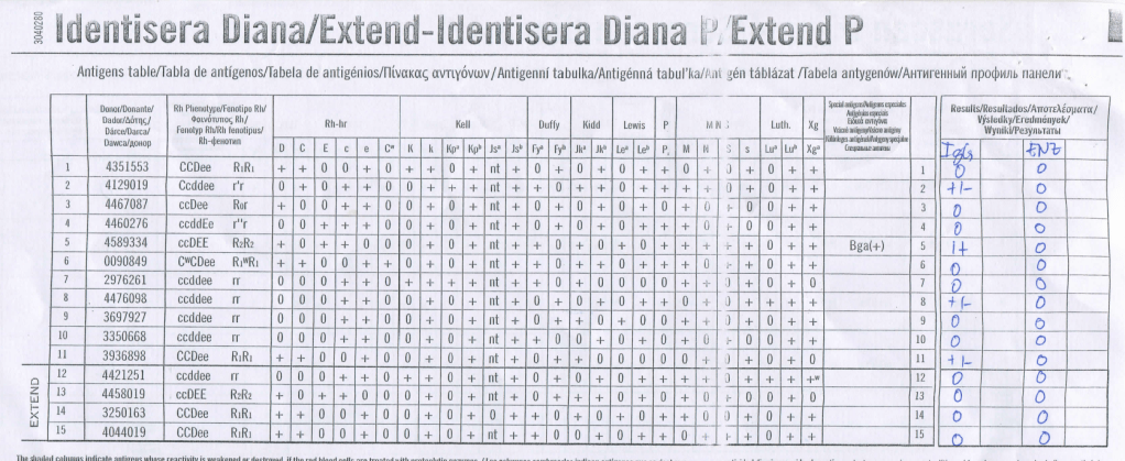
This is an example of a nonspecific reagent using whole molecule IgG AHG:

Here is the same sample using gamma-heavy-chain-specific AHG:

4/10/20

Projective Exercise 8 Solution: D-positive with anti-D

Some Possible Explanations:

Always review the transfusion history of all component types, medication history, and the clinical history!! Start with this first.

  1. Receipt of plasma with anti-D (RhIG, IVIG, etc.)–passive antibodies
  2. Partial D with anti-D:
    1. Partial or mosaic D patient who received D positive RBCs and made anti-D directed against its missing epitopes
  3. Anti-G:
    1. Not all anti-G is anti-C and anti-D:  It is really a separate specificity.  It is possible that anti-G may be made even though the patient is C-positive.
  4. Anti-LW:
    1. However, it is unlikely to show such strong reactions

Can you think of other explanations?

3/10/20

Processes and Software Building 49: Donor Automated ABO Typing

For donor ABO typing, I always had both automated and manual methods set up on the blood bank computer system Medinfo Hematos IIG.  The automated method had a bidirectional interface between Medinfo and the instrument.   Medinfo did not need a separate middleware.  A truth table was prepared for acceptable results for automatic interpretation.  Other results had to be manually interpreted by someone with the appropriate security level.

The manufacturer’s recommendations for the particular reagents in use were strictly followed.  One used the range {0, 1, 2, 3, 4} as acceptable.  Another used {0, 2, 3, 4}.  Controls were included.  Refer to the following flow diagram.

Most importantly, Medinfo could be configured for any set of reagent values.

2/10/20