Nonspecific Antibodies: My Approach

Anyone reviewing antibody panels, especially in the Middle East/Gulf region, encounters many panels for which no antibody specificity is identified.  As a transfusion medicine physician who often got called during the night for release of RBCs for patients with “nonspecific” pattern, this was a big headache.

Is it “nonspecific” because there isn’t a clinically significant antibody OR the technologist did not perform the testing or its interpretation correctly?  Does it need further testing?  Do I release blood components at this time?

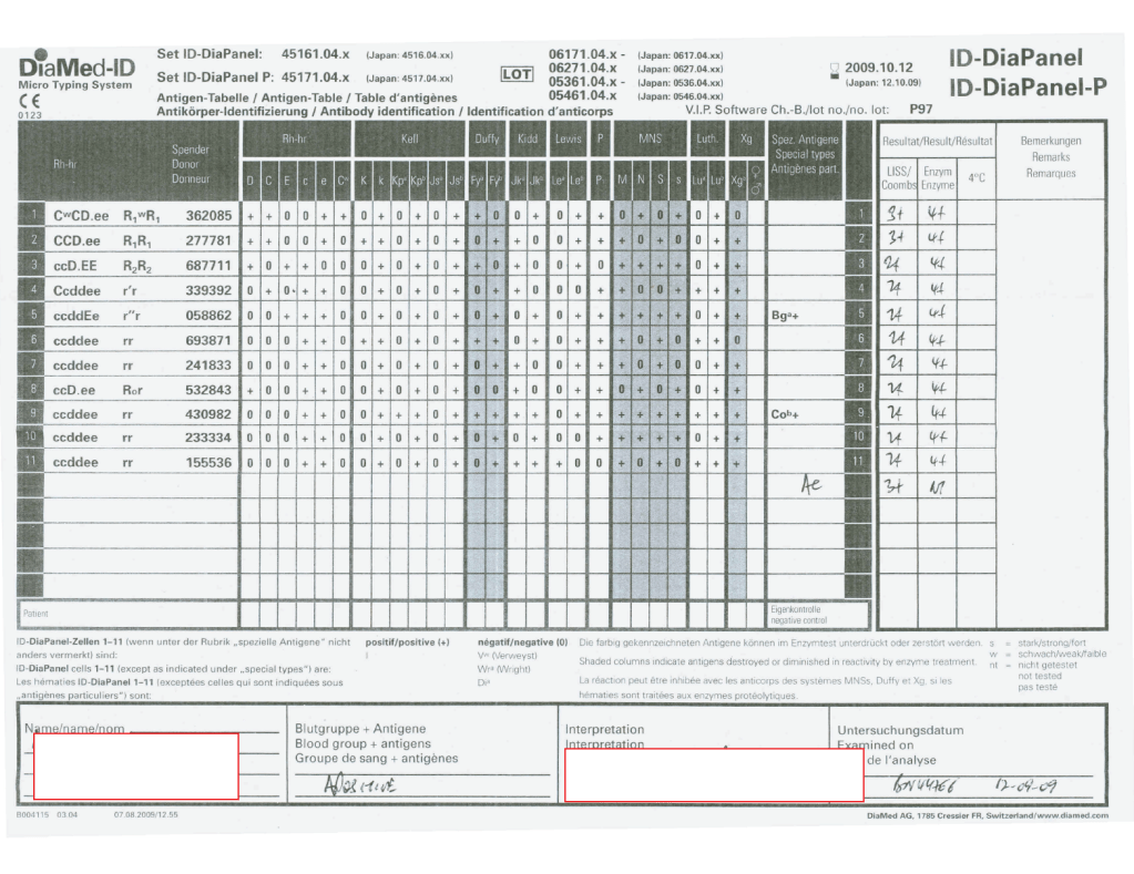
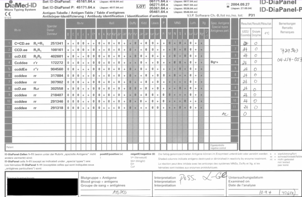
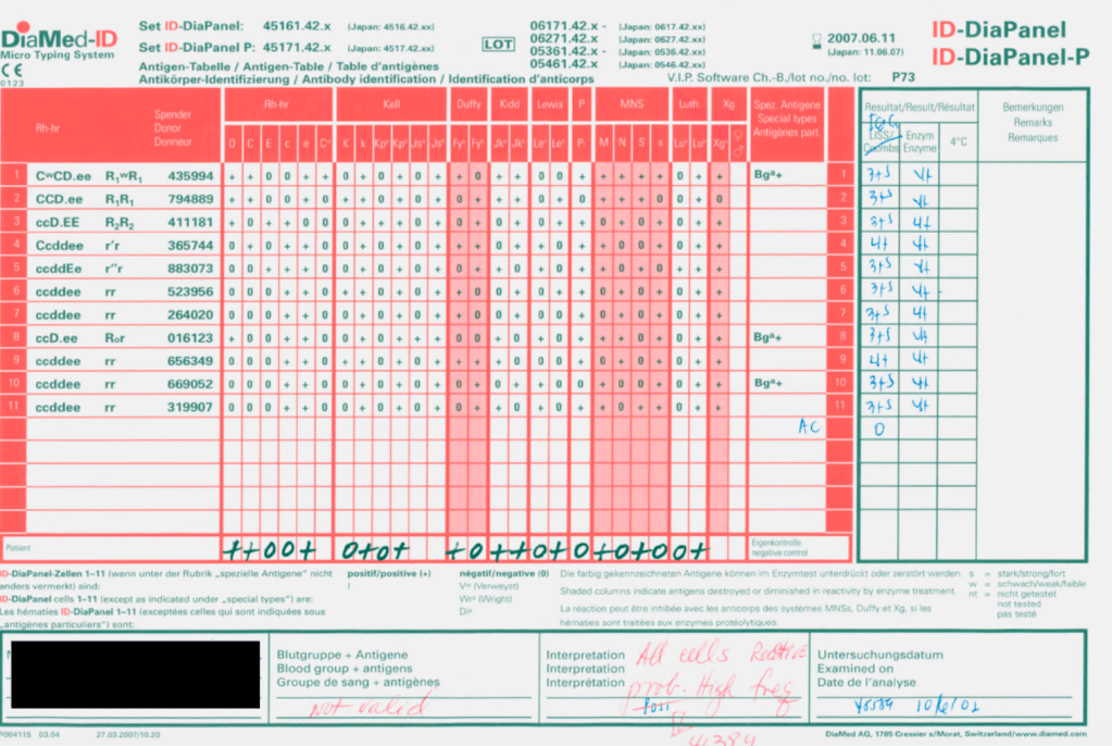
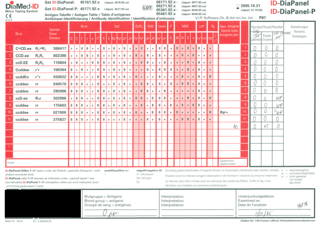
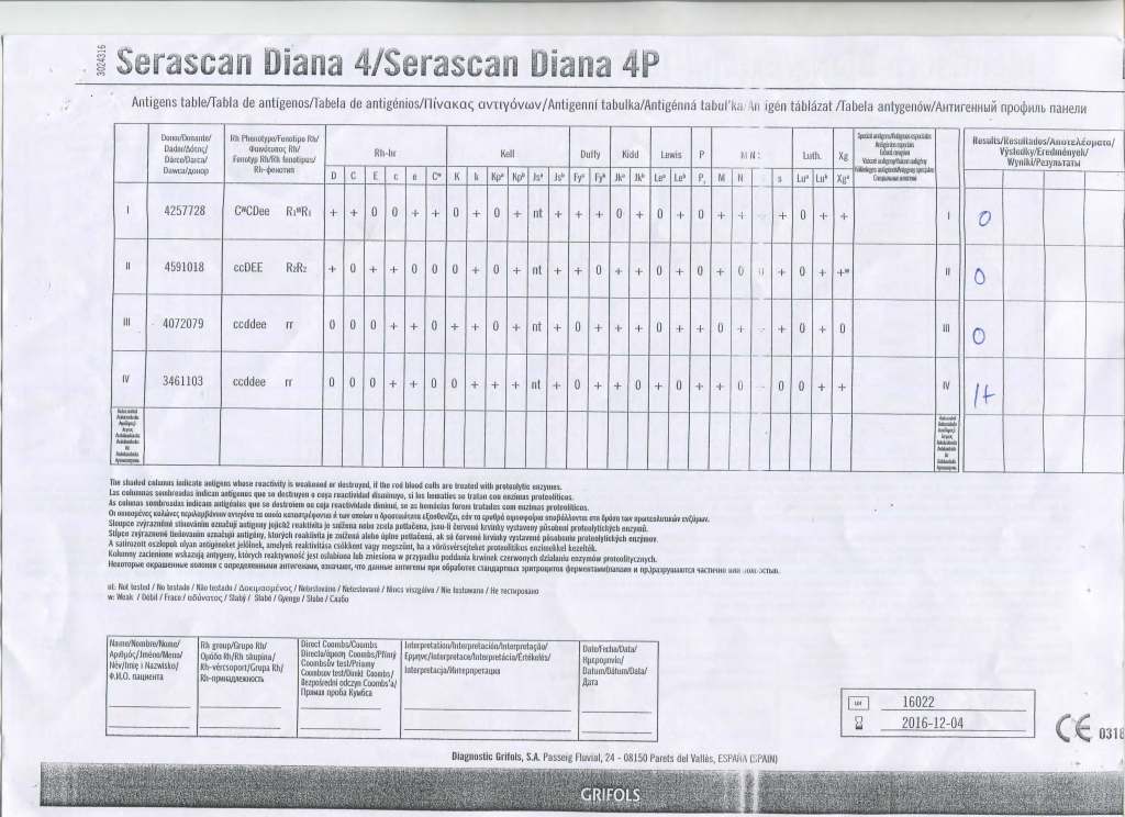
I have attached an example of an antibody panel that has a few weak to 1+ reactions using a gamma-whole-molecule IgG AHG reagent.  Notice that they are enzyme-labile.

I have worked in this region both before and after gel/glass bead technology was introduced.  In the good old days, I formerly only used a gamma heavy-chain specific AHG in tubes with LISS.  Nonspecific reactions were few.

When we adopted gel and at some sites glass bead columns, there was only a choice between polyspecific AHG and whole-molecule IgG AHG.  The rate of non-specificity soared from less than 10% to over 30% of panels.  The reactions were reproducible over multiple technologists at multiple sites.

When I repeated the same specimen against a manual tube panel using gamma-heavy-chain specific AHG, the reactions disappeared in the majority of the cases.  Why?

Heavy chain specific reagents do not detect light chains.  Light chains are the same in IgM and IgG antibodies.  Both polyspecific and whole molecule IgG AHG detect light chains.  Thus, high-thermal-amplitude cold antibodies may be detected with the latter but are unlikely to react with heavy chain specific reagents.

When I used manual tube reagents, I would have missed these reactions, but I do not recall this having clinical consequences except in very rare circumstances (refer to my previous post about the anti-Jka only detectable with polyspecific reagents—I have seen 3 in 30 years).

I have asked various gel/glass bead manufacturers to provide me with heavy-chain gamma-specific AHG, but none have agreed.

Normally, for automation, I use IgG-whole molecule AHG.  If the reactions are nonspecific, we repeat the testing manually using the gamma-heavy-chain-specific AHG.  If negative, I ignore the nonspecific reactions and use the same gamma-heavy-chain-specific AHG for full antiglobulin-phase crossmatching.

In general, with nonspecific reactions, I recommend the following:

  1. Always do enzyme panels, sometimes with both papain and ficin reagents:  many Rh antibodies are optimally detected only at enzyme (example:  R1R1 with apparent anti-E at AHG but the anti-c only seen at enzyme)
  2. Perform an extended Rh/Kell/Duffy/Kidd/Kell/MNSs/P1 phenotype and specifically check for those negative typing results AND for dosage (could the antibody only be detected in homozygous cells like many anti-M are?).
  3. Perform classical room temperature, 37C, and finally AHG phase testing.  Routinely I do not do this since antibodies not detected at 37C are unlikely to be clinically significant.  Sometimes, the AHG phase reactivity is a cold antibody of high-thermal amplitude.
  4. If the Jka or Jkb antigen typings are negative, repeat using a polyspecific AHG reagent.
  5. Use additional panels from multiple manufacturers.  Some reagents detect more nonspecific reactions than others.
  6. Try other potentiators than LISS such as PEG.
  7. Check the outdate of the panel and reagents:  if less than 1 week remaining, consider repeating with fresh reagents and getting a new patient sample.

Finally, if you still cannot define the specificity, consider repeating after several days.  Maybe it is a newly emerging or an anamnestic response.

I emphasize as a physician, I do not care to see all possible antibodies present in the specimen but rather only those likely to be clinically significant.

Processes and Software Building 9: Enforcing GMP

Enforcing Good Manufacturing Process Through A Dedicated Blood Bank Software

Blood components are a drug and like medications must be consistently produced and follow Good Manufacturing Practices GMP.  The following system that I set up for HMC Doha Qatar blood collection and processing is an example of the impact of Medinfo donor software on enhancing our safety and GMP compliance.  In earlier posts, I provided the Medinfo flowcharts for these processes.

This is an outline of the processes I built in conjunction with the Medinfo software engineers:

  1. Registration:
    1. Read the barcode on the specified picture ID (usually a Qatar Residency/Citizen card).
      1. Retrieve the prospective donor’s demographics in English and Arabic from the Ministry of Interior.
      2. Check the donor deferral database (Qatar has only one), defer if contraindicated according to the rules built into Medinfo.
    2. Choose the type of donation (apheresis or whole blood;  volunteer, directed, or autologous).
    3. Print a consent form and ISBT specimen labels for the donation.
  2. Pre-Collection Screening:
    1. Perform the donor questionnaire on-line in English or Arabic:  this has contingent fields and could exceed 60 questions depending on the answers provided.
    2. Proceed to donor physical exam (vital signs and arm check).
  3. Collection:
    1. Collect the whole blood or apheresis component and specimen tubes:  determine if the collection meets the volume requirement and time limit.
    2. Send the specimens for donor marker and donor immunohematology testing.
    3. Send the raw components for processing.
  4. Donor Marker Testing:
    1. Perform NAT, EIA, and LIA marker testing according to algorithms defined in Medinfo.
    2. Perform follow-up reflex marker testing according to Medinfo criteria.
  5. Component Processing:
    1. Process the raw whole blood in the Reveos machine into PRBCs, leukodepleted plasma, and buffy coat platelets.
    2. Filter/leukodeplete the RBCs.
    3. If marker test results pass:
      1. Pool the buffy coat platelets according to the platelet yield index for a yield of 2.4E11/dose.
      2. Add platelet additive solution PAS and pathogen inactivate (Mirasol).
      3. Pathogen inactivate the whole-blood-derived plasma.
      4. Divide the apheresis plasma into 200-250 ml aliquots and pathogen inactivate.
    4. Divide the apheresis platelets to provide a yield of 2.4E11 in each dose, then pathogen-inactivate (PAS had been added at the time of the apheresis collection).
  6. Donor Immunohematology Testing:
    1. Perform ABO/D testing and antibody screen (identification if positive):
    2. If antibody screening positive, discard the component.
      1. If ABO discrepancy, send for manual review and approval, otherwise discard.
  7. Labelling, Storage, and Transfer:
    1. If all criteria were met, attach the final ISBT label (this can only be printed based on the acceptance of each component).
    2. Place the components into storage (37C, 1-6C, or <= minus 18C).
    3. Distribute to the hospital blood banks using Inter-Depot Transfer function.

I emphasize that only if all criteria across all areas pass is the final ISBT label printed.  Medinfo is not a label printing program.  It enforces the rules ruthlessly.  My technical staff tell me that it is merciless—as it should be for patient safety.

Attachments:  None—please refer to earlier posts regarding collection, processing, donor testing, and inter-depot transfer.

28/6/20

The Devil is in the Details: Three Antibody Panels for Illustration

As a Transfusion Medicine physician, I reviewed all antibody workups.  Here are three different panels, all appearing to be panreactive,  at AHG phase.  Note the differences:

  1. Autocontrol is positive, enzyme is panreactive and enhanced
  2. Autocontrol is negative, enzyme phase shows no reactions
  3. Autocontrol is negative, enzyme phase is panreactive enhanced

In the middle of the night, if I am called to select blood, I always keep these patterns first and foremost in my mind.

Case 1:

If the autocontrol is positive at about the same strength as the panreactivity.  I can state that this is probably a warm autoimmune pattern WAIHA (although I cannot rule out underlying clinically significant alloantibodies obscured by this pattern and need to do autologous auto-absorption ZZAP).  Both warm autoimmune antibodies and certain drug reactions may cause this pattern.  Use least-incompatible crossmatch matching any significant specificities you found by ZZAP and observe the patient closely throughout transfusion.

Case 2:

If the autocontrol is negative and all reactions are enzyme-labile—and if you live in the Middle East/Gulf region, then you have a presumptive anti-Ge2.  You can safely ignore this antibody and release least-incompatible crossmatch RBCs, regardless of the strength of the reactions.

Case 3:

If the autocontrol is negative and the reactions are unchanged or enhanced by enzyme, BE AFRAID, VERY AFRAID!!  This is an antibody to a high-incidence antigen.  These can be very dangerous.  In the Middle East/Gulf region, consider anti-H, anti-k (cellano), anti-Kpb, anti-PP1Pk, and rare antibodies to the MNSs such as anti-U or other MN system deletions.  There are many other possibilities, e.g. anti-Fy3.

You need to perform extended antigen typing across the major Rh antigen, Kell, Duffy, Kidd, MNSs, P systems (at least P1):

  1. Run H lectin to rule out Bombay Oh or Parabombay.
  2. Some Rh system deletions and Rh null show pan-Rh reactivity—check D, C, c, E, e typings.
  3. Anti-k (cellano) will be suggested by k-negative phenotype.
  4. Unusual, weak or absent reactions with M and N reagents suggest something like En(a)-negative or similar.
  5. Absent P1 with no other findings, you must rule out anti-Tja (anti-PP1Pk).

I have seen all of these specificities during my time in the Middle East.  All of these antibodies can be clinically significant and often life-threatening.

Conclusion:  NEVER NEGLECT TO REVIEW THE AUTOCONTROL!!!

Processes and Software Building 8: Reveos and Mirasol

Automated Component Processing:  Reveos and Mirasol Pathogen-Inactivation

The production instruments have more complicated interfaces than the testing equipment discussed in the previous post:

In the collection area (on-site or remote), the cvolume of the whole blood and collection time are recorded in Medinfo and based on the rules, production may only occur within specified volume and collection time.  Otherwise, Medinfo will block further processing.

The ISBT unit number of the whole blood units are read by the Reveos.  Only those units passing the collection criteria will proceed to separation.

In about 20 minutes, the Reveos machine will simultaneously process four units of whole blood into packed RBCs, leukodepleted plasma, buffy coat platelets, and residual buffy coat.  The volumes of the RBCs, plasma, and platelets are recorded in Reveos.  For the platelets, the platelet yield index is also provided.

Within Medinfo, these parameters are compared to criteria of acceptability according to the manufacturer.  Volumes for the platelets and plasma must be within certain ranges to permit pooling and pathogen inactivation and additive solution.  Medinfo will not permit these subsequent procedures if the values are out of range and the intermediate components will be discarded.

Here is a sample of Reveos acceptable ranges for component volumes:

E4207 – Whole Blood CPD 450 mL

Volume Consequences

< 400 mL Discard

400 – 500 mL OK

> 500 mL Discard

E5259 – Leukodepleted Packed Red Blood Cells

Volume Consequences

< 230 mL Discard

230 – 330 mL OK

> 330 mL Discard

E2807 – Platelets Concentrate 20-24°C

Volume Consequences

< 20 mL Discard

20 – 55 mL OK

> 55 mL Manual decision

E2555 – FP24:  Plasma Frozen <= 24h

Volume Consequences

< 170 mL Discard

170 – 360 mL OK

> 360 mL Manual decision

All these production parameters are permanently stored in Medinfo as part of the production record of that unit.  The actual location (bucket) of the whole blood unit in the Reveos is also available.

RBCs are manually leukodepleted and the final volumes recorded in Medinfo based on weight.  Based on the platelet yield index, platelets are pooled and the final volume recorded.   Those permissible volumes are next treated with platelet additive solution PAS and then pathogen inactivated.  The acceptable volumes are based on the process used, e.g. platelets in plasma versus platelets in PAS.

How a sophisticated blood bank software like Medinfo enforces good manufacturing process at all stage of production will be a future topic.

To Be Continued:

26/6/20

Nonspecific Reactions, Reagents Near Expiration

Whenever I had a “nonspecific” antibody, I had to first rule out issues with the reagents themselves. The following example shows weak to 2+ reactions in the panel cells and autocontrol.

The variability in the reactions made me initially uncomfortable about called this WAIHA. I then checked the panel details: the testing was done only six days before the panel outdate.

I told my staff to repeat the workup with the new panel expiring five weeks later. The difference is astounding!!

Remember: if you work in the Middle East, the environmental conditions can be extreme in summer (>50C). Do you know how your reagents were handling during transport?

My advice: if you are concerned there is a clinically significant antibody but cannot discern it, consider repeating the workup using fresh reagents.

Processes and Software Building 7: Interfaces 2

Blood Bank instruments may perform tests and release test results in a numerical or alphanumeric format or both.  For example, nucleic acid and enzyme immunoassay may release a qualitative result (e.g. positive, reactive, borderline/grayzone, negative, nonreactive).  Alternatively, the machine may release the signal to cutoff ratio (S/CO) as a numeric result.

Blood bank software may use either kind of result on which to base interpretative rules for acceptability of the donor.  The qualitative result criteria are based on the quantitative SC/O but the equipment automatically interprets this.  The S/CO ratio of 1 is the cut-off point.  Thus a value of 0.99 is negative and the value 1.01 is positive.  But is it really so clear-cut since the difference between the two is so small?  Thus, some people have added the term grayzone for values close to but below the cutoff.  Could a value of 0.95 be an early infection?

I personally prefer to see the actual cutoff but use the manufacturer’s criteria for interpretation.  As a physician, it is good to review the S/CO on serial exams.  If a borderline or grayzone result becomes positive, then perhaps the original result indicated early infection.  The question still remains, what is the grayzone?  0.95 to 0.99, 0.90 to 0.99, etc.  Some accrediting schema have not used grayzone for interpretation.

With Medinfo’s blood bank software, I could chose either option or both—or at least store the S/CO as a nonreported result for subsequent review.  I could even chose, test by test, in a series between reporting either S/CO or the qualitative result.

Semiquantitative results, e.g. in {0, 1+, 2+, 3+, 4+} are qualitative and could also include mixed field (mf) and hemolyzed (h).  I showed examples of this with ABO/D antigen typing in a previous post—see attachment.

On the contrary, the results from blood production equipment may include parameters such as time of preparation, original volume, final volumes for each component, platelet yield index as an indirect measure of platelet count.  When there is pooling, the final total volume is critical to determine if pathogen-inactivation procedures and platelet additive solution can be used.  This is a much more complicated interface.

The blood production equipment interface issues will be considered in a future post.

Attachment:

ABO/D sample typing process in Medinfo

To Be Continued:

24/6/20

Case Report: Unusual Antibody Anti-Jka

This is a rare anti-Jka that only reacts with polyspecific AHG and only in Jka+ homozygous reagent cells. In the past three decades in the Middle East, I have only seen three of these.

Almost all anti-Jka and anti-Jkb antibodies in my experience can be detected using monospecific, gamma-heavy-chain AHG reagents (I personally prefer this latter reagent to avoid cold antibody interference.)

If you have a nonspecific antibody, never discount this possibility and then run polyspecific reagents and check for dosage. I also recommend extended antigen typing (e.g. Diamed/Biorad Profile Cards 1-2-3).

Processes and Software Building 6: Interfaces–General Considerations

When buying equipment while planning/implementing new laboratory software, I originally had a rule not to purchase anything that the vendor did not have a ready interface.  Even that was not so clear since some vendors had interfaces listed as alpha, beta, and completed.

Could you use an alpha or beta interface?  Was it safe for patient care?  What was the development cycle for new interfaces with your vendor—months, years?

Even if the vendor had a completed interface?  How “complete” was it?  Did it accept all data from the machine?  Did the data stream require reformatting?  Who would write the transformational script?

Even if the vendor could support it, could your local IT organization and the local agent’s IT staff do it?  I had plenty of headaches over this.  The best equipment with the best interface that the local agent could not support was worthless to me.

Some finished interfaces took months to install because of connectivity issues?  What version of the operating system was used?  Was it secure?  Did our IT department accept that version (e.g. Windows 7) and the provided malware protection?  I have seen malware spread across a network from the interface software installed by the vendor, threatening the entire corporate system. 

Did the solution require middleware?  What were the implications of having middleware and its affect on the main software program, especially at the time of its upgrade or the main software?

I have seen vendors using Windows 2000 for their interface software as late as 2017.  It was difficult for some of them to update to current, more secure versions.  Anyway, our corporate IT department gave them all a deadline to update to the current operating system—they all complied or risked losing all connectivity to the network.

Almost every instrument vendor has told me that they can communicate with my laboratory system.  I guess that is true:  one talks in Russian and the other Sanskrit—they do communicate but is it effective?  Talking is not necessarily communication!

I remember one open EIA machine that had a TCP/IP port but it was not functional by the standard protocols.  One had to emulate a serial port to get some rudimentary communication.  The port’s light blinked, however.  I never imagined that someone would put a nonfunctional port as a mere decoration.

On the other hand, I have had excellent experience with another software vendor Medinfo.  Even if the vendor did not have the interface developed, they could build it from scratch in a few weeks.  Paradoxically, it was faster for these new interfaces than some so-called already interfaces.

I must emphasize:  This is a collaborative team effort between the blood bank information system, software vendor, instrument vendor, and your institution’s IT staff.  There must be excellent cooperation between them for a successful result.

When installing the Medinfo Hematos IIG software, many of our most important interfaces (the Terumo mixed shaker, Trima, Reveos and its predecessor Atreus, Mirasol illuminator) had no developed interfaces when we started,  This was a risk;  but actually those interfaces were developed in a few weeks and fully functional.  In fact, we were the first site in the world to have those interfaces working—and without any Middleware.

In general, the blood bank software vendor installed the completed interface and did some low-level testing.  Then, my blood bank computer team did the testing.  The final responsibility for testing and acceptance was with the end-user blood bank team.

I am attaching a copy of our Abbott Architect interface as updated a few years ago.  Again, here I wrote the validation protocol and assigned the tasks to the Medinfo Super Users.  To perform this EIA testing, we still had to register donors and collect and then export the specimens to the donor marker testing laboratory before the actual interface testing could begin.  This was all done in a special test domain separate from the production domain.

I made the validation criteria and reviewed all data as Division Head of Laboratory Information Systems.  Representative screen shots were made.  All data was sent to me.  My final acceptance was required before the interface could be activated.

Automated component processing (Reveos) and component modification are more complicated and will be covered in a future post.

Attachment:

Abbott Architect Interface

To Be Continued:

24/6/20

Processes and Software Building 5: Processes

Processes and Software Building—Part Five

This post is mainly on building processes for a non-turnkey system such as the Medinfo Hematos IIG software that I have worked with in several countries, but there will be a few words about turnkey systems for general laboratories.

This has been a collaborative effort between the software vendor’s engineers, my Super Users, and myself.  This pluralistic approach has been most productive.

A turnkey system has pretty much already defined most of the basic processes—those have been specifically approved by a regulatory agency such as US FDA.  There is little customization except formatting screen and reports.  Instrument interfaces are also mainly predefined.  This requires much less thought and planning than a custom-built system designed on the sites actual workflows, but it can be an exercise of putting a round peg in a square hole.  You don’t always get what you want.

In the locations where I collaborated in setting up the Medinfo Hematos IIG program, we did not follow US FDA but mainly the Council of Europe CE standards since this was much more customizable and applicable to our needs (KSA and Qatar).  We could modify and add additional criteria specific to our country and region (e.g. rules for donor qualification for local pathogens).  This has always been my preferred approach.

Start with a frame of reference (CE) and then try to optimize it for our local needs.  Unfortunately for blood banking, FDA has many fewer approved options than other regions, including in the preparation of blood components (e.g. prohibiting the use of pooled buffy coat platelets, automated blood component production such as Reveos, and use of world-class pathogen-inactivation technologies) such as Mirasol.

If you invested the time to make a detailed workflow across all processes and tests, much of this can be readily translated into the software processes, but first you must study the flows and determine where you can optimize them.  This requires that you study the options in the new software to see what you can use best.

I always liked Occam’s Razor, i.e. “ntia non sunt multiplicanda praeter necessitatem,”—the simpler the better as long as it meets your needs.  If the manual processes are working well and can be translated into the new system, do so.  If they need changes for optimization, then do so only if necessary.

Most of my career has been spent overseas with staff from many different countries and backgrounds, most of whom were not native in English.  The wording of the processes is very important.  Think of the additional obstacle of working with a complicated software in your non-mother tongue!  Also consider the differences between American English, British English, and international English.  I always made the Super Users read my proposed specifications and then asked them to repeat what I wrote/said.

There were many surprises discovered.  I think of the Aesop’s fable about the mother who gave birth to an ugly baby looking like a monkey.  Still, to that mother her baby was the most beautiful baby and she entered him into a beauty contest.  In other words, to the mother her child is perfect!!

It is most important to use the manufacturer’s recommendations to build tests and for the special automated processing and pathogen-inactivation processes.  For example, we had multiple ABO and D typing tests—they did not necessarily agree on what were acceptable results for automated release of results.  The same is true for many other tests. Use the manufacturer’s recommendations.

Example:  One method for Rh(D) typing stated that only results in {0, 2+, 3+, 4+} were acceptable—all other results required manual review and/or additional testing.  Another only accepted results in {0,3,4}.  Thus we had to build separate D typing processes for each methodology.

If we changed equipment at one site to that used at another site, we didn’t have to modify our software to accommodate this.  Even if you didn’t have the equipment or reagents at one site, you could always build it into the system and not activate the settings until needed.

Another consideration is whether to offer all the processes globally or restricted to one site.  I favor allowing access to all methodologies at all sites—in case of a disaster where tests had to performed at another site.   This means that if you send an order over an interface from the hospital system to the blood bank system, that at the receiving (blood bank) end, you would choose which methodology to use, i.e. it is not a one-to-one but rather a one to many mapping.

Finally, the issue of middleware.  Many instruments offer this, but one faces the problem about support and regression errors when you either update the middleware software or the blood bank computer software.  Medinfo itself could serve as the middleware so there was less chance of errors when updating the software.  In fact, I have never used any middleware when using Medinfo.

Instrument interfaces will be a future topic.

To Be Continued:

23/6/20