Therapeutic phlebotomies TP, like regular whole blood donation WBD, both require collection of whole blood into a blood bag set. In whole blood donation, we ask two questions during the process:
- Is it safe for the donor to donate?
- Is it safe for the collected unit or its processed components to be given to recipients?
For TP, we only have to consider the first question so the process must ensure the patient/donor’s safety. We do not have to concern ourselves with the use of the collected product—it will be discarded.
Since the donor collection processes of both TP and WBD are similar, why couldn’t we use the blood bank computer software to document the TP procedures? The process is a subset of normal WB donation. On this basis, I make suggestions on using the donor module to document the TP process. It is basically a truncated version of blood donation process in the Medinfo Hematos IIG system:
- Registration
- Donor Safety
- Vital Signs
- Hemoglobin Determination
- Blood Collection Data
- Adverse Effect Reporting
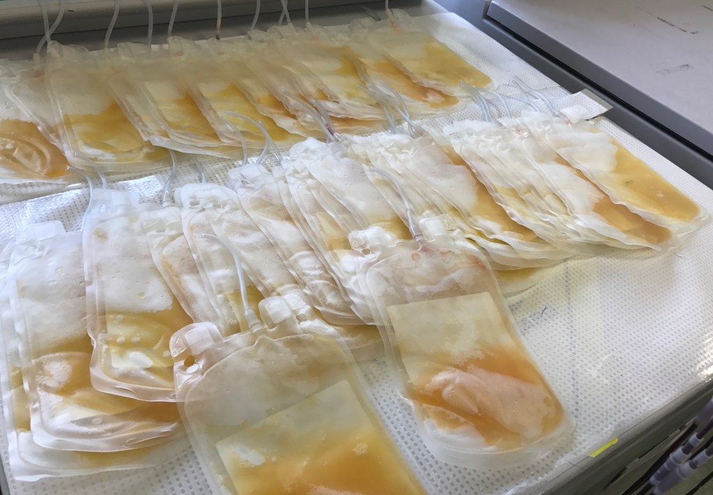
- Discard of Unit
- Documentation of Physician’s Order and Transfusion Medicine Physicians Acceptance
Registration: Positive patient identification can be made through the donor registration process; force selection of an inexpensive bag type (not the Reveos set) for this purpose.
Donor Safety: Perform a modified, shortened donor questionnaire covering the medical history and medications is used. Confirm that the patient has had food and drink before donating. Require a waiting period of 24 hours before the next procedure.
Vital Signs and Weight: Measure weight plus BP, pulse, temperature, and respiratory rate as well as inspect the arm for scarring before procedure. Allow repeat vital signs monitoring after the procedure if requested by the transfusion medicine physician.
Hemoglobin Determination: Allow acceptable Hgb >= 11 g/dl or >33% hematocrit
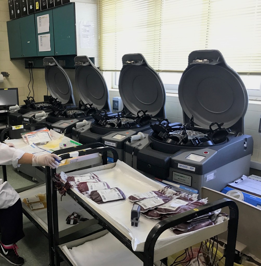
Blood Collection: Use the same process for the mixer-shakers but the amount collected can range up to 500 ml with amounts <405 ml acceptable for small patients
Adverse Effect Reporting: The complications of TP collection are the same as WBD. Use the same system as for WBD.
Discard of the Unit: Print discard label and quarantine of the ISBT unit number in system (so that it cannot be used for transfusion).
Documentation of Order: Create separate fields for the ordering physician and for the approving transfusion medicine physician. Capture scan of paper orders and incorporate into the TP computer encounter.
Other Considerations: In high-risk cases, e.g. with pre-existing cardiovascular, pulmonary, or cerebrovascular disease, one could consider using a remote monitoring device such as the Umana T1 device to record vital signs, EKG, and oxygen saturation that can continuously record these parameters and trigger user-definable alarms during the process and afterwards if desired. The data can be incorporated into the blood bank computer encounter.