Autologous Transfusion and Responsibilities

This is the process I developed for HMC Doha. The Medical Director (here Head, Transfusion Medicine HTM) is actively involved in the development of policies, processes, and procedures for ALL types of autologous donation in conjunction with the National Transfusion Committee NTC.

  1. Predeposit:  Directly under the control of the HTM for all aspects:  policies, procedures, and direct performance of the procedures, including annual review of criteria
  2. Perioperative:  HTM involved in conjunction with Surgery and Anesthesia through the NTC.
  3. Intraoperative:  HTM involved in conjunction with Surgery and Anesthesia through the NTC.
  4. Postoperative:  HTM involved in conjunction with Surgery and Anesthesia through the NTC.

Background:

There are four basic types of autologous transfusion:  preoperative, perioperative hemodilution, intraoperative, and postoperative drainage/collection.  The use of all of the above techniques can significantly decrease the need for homologous blood and as an added benefit reduce the risk of the disease transmission and immunosuppressive effects of such homologous transfusions.

Preoperative collection can make available packed red blood cells, whole blood, platelets, FFP, and/or cryoprecipitate.  However, at most two units of blood per week can be collected.  RBC’s can be stored for up to 42 days in the liquid state, frozen RBC’s up to ten years, platelets up to five days, and fresh frozen plasma and cryoprecipitate up to one year.  The last collection cannot be less than 72 hours prior to the surgery time.  Units can be collected as long as the patient’s hematocrit remains above 33%.  Supplemental iron and erythropoietin can increase the number of units harvested.  The biggest obstacle to using this service is the coordination of the patient scheduling for this procedure.  The blood bank does not have the resources to prospectively analyze the surgical scheduling and make the various appointments, contact the attending physician, etc.  Thus, this service is vastly underutilized.

PHD or Perioperative hemodilution (also called acute normovolemic hemodilution) is useful in cases when the anticipated blood loss is at least one liter and the initial hematocrit is at least 34%.  This includes essentially all types of surgery, but in particular cardiac, vascular, orthopedic, and urologic cases.  The patient’s hematocrit Hct. is lowered to the range of 20-25% and the blood is replaced by crystalloid in a ratio of 3:1–i.e. three times as much fluid as blood, or in the case of colloid replacement, a 1:1 ratio of colloid plus 0.5 to 1.0 ml. of crystalloid.  Crystalloid has the advantage of being readily removed by diuretic use.  However, this technique should not be undertaken when vascular access is inadequate or appropriate monitoring devices are lacking.  The physician performing PHD must be familiar with the compensatory mechanisms normally invoked when the hemoglobin is acutely lowered.

Another new twist to PHD is the perioperative collection of platelets by a special attachment to a cell-saving machine.  This could allow collection of a typical apheresis load, about 6 to 10 units of fresh platelets for potential use.  There are currently studies underway to determine if this has particular clinical advantages to warrant the additional cost.

Intraoperative salvage may be performed with a number of canister or automated devices.  The latter is usually used when there are large volumes (usually 3 or more units) of blood to be salvaged.  Depending on the body site, the recovered material is at least filtered and may or may not be washed.  Care must be taken to collect the blood at a low suction rate and with minimal turbulence to minimize hemolysis.

Postoperative drainage collection of certain sites such as post-knee replacement surgery or chest wounds involves a canister collection device.  This blood may or may not be filtered before reinfusion.

Note that perioperative and intraoperative material can only be transfused up to six or eight hours at room temperature or 24 hours if refrigerated at 1-6 degrees (depending on the method used) post collection to minimize the risk of infection.  Intraoperative collection is usually contraindicated in cases of cancer and if the bowel has been violated.

Other Issues:

The transfusion criteria for autologous blood is the same as for allogeneic units. If you wouldn’t transfuse if no autologous blood were available, you shouldn’t transfuse because you have it!

The same compatibility testing algorithm applies both the autologous and allogeneic units.

Policy:

  1. Scope:
    1. Predeposit collection of Whole Blood/RBCs and plasma is under the authority of Transfusion Medicine.
    2. Perioperative hemodilution, intraoperative cell salvage, and postoperative drainage collection is under the authority of the National Transfusion Committee in conjunction with the Departments of Surgery and Anesthesia.
      1. Head, Transfusion Medicine will liaise with the clinical departments as needed.
      2. Transfusion Medicine may provide blood bags for perioperative hemodilution upon request.
    3. Transfusion Medicine does not receive autologous collections—perioperative, intraoperative, or postoperative drainage collection.
  2. Processes directly under Transfusion Medicine authority:
    1. Autologous collection of whole blood/RBCs (predeposit) for elective surgeries will be considered especially if the patient has a dangerous antibody for which antigen-matched units cannot be easily obtained (e.g. anti-k (cellano), anti-PP1Pk (anti-Tja), anti-H (Bombay and Para-Bombay phenotypes).
    2. Autologous collection of plasma may be considered for patients with IgA deficiency with documented specific anti-IgA antibodies.
    3. Autologous collection of platelets may be considered for patients with anti-platelet antibodies and platelet refractoriness.
    4. Other requests will be reviewed by the Head, Transfusion Medicine or designate.
    5. The final decision to proceed with items 2.1 and 2.2 will be made by the Head, Transfusion Medicine or his designate.
  3. Process for Transfusion Medicine Autologous Procedures
    1. The requesting physician shall provide a written order to the Blood Donor Center.
    2. The request will be reviewed by a transfusion medicine physician.
    3. If rejected, the requesting physician will be notified with the reason for the rejection.
    4. If approved, the donor shall be screened by the usual donation process except:
      1. Hgb >= 11 g/dl will be acceptable for whole blood collection.
      2. Females may also donate autologous plasma.
      3. The last autologous donation will be at least 72 hours before the elective procedure.
    5. Marker testing:  Components from autologous donors with confirmed positive cases of HBV, HCV, HIV, syphilis, malaria, or HTLV infection will NOT be used for autologous donation and will be destroyed.
    6. Computer:  Autologous collections will be entered as specifically as such in the Medinfo  Hematos IIG blood bank computer system and be labeled as autologous in their corresponding ISBT labels.
    7. Crossover of autologous units requires review and approval by a transfusion medicine physician–only those cases where the donor met the standard donor criteria will be considered.
    8. The transfusion criteria for autologous units shall be the same as for homologous blood.
    9. Both autologous and allogeneic units will follow the same compatibility testing algorithm.

References:

  1. Standards for Blood Banks and Transfusion Services, 29th Edition, AABB, Bethesda, MD, USA, 2014
  2. TRM.41600 CAP Checklist Standard, 2015

Processes and Software Building 13: Validation of Electronic Crossmatch

The electronic/computer crossmatch is vital to the hospital blood bank’s operation.  It allows us to reduce unnecessary work and redeploy a very limited, precious workforce.  As demonstrated in the previous post, it has many rules for current and previous testing and historical transfusion records.

This is NOT a one-time validation:  it must be repeated to check for regression errors when updating the software version, regardless if there are any changes to its logic.  It required many Super-Users to perform:

  1. A test environment distinct from the live was used.
  2. Patients had to be created and specific test results entered to match the algorithm below.
  3. Donors had to be created and components made (including testing, production, release, inter-depot transfer)
  4. Hospital blood bank had to receive the created units and enter them into active stock (and repeat the ABO/D testing to do this)

This is a sample validation plan created by me and used at HMC Doha for the Medinfo Hematos IIG software:

  1. Special Electronic Crossmatch Testing
    1. Make 100 patients:
    2. 60 patients matching electronic crossmatching criteria:
      1. 2 ABO/D determinations, one current within 72 hours
      2. Second determination from history or second current sample
      3. No ABO/D typing discrepancies
      4. Negative antibody screen
      5. No history of antibodies
    3. 40 patients with contraindications to electronic crossmatch:
      1. 10 with history of antibody
      2. 10 with current antibody
      3. 10 without 2 ABO/D determinations
      4. 10 with emergency release
    4. Try to release RBC unit without further testing in each case.

Super Users had to capture screen shots of the transactions and send them to me as Division Head, Transfusion Medicine/LIS for review and acceptance.

Criteria for Acceptability:

  1. All tested processes must complete without error, including recognition of ABO/D discrepancies as such.
  2. System only allows computer release (electronic crossmatch) in those cases meeting computer-crossmatch criteria as shown in the following interim policy.

INTERIM POLICY:  ELECTRONIC (COMPUTER) CROSSMATCH

Revised Date:  1/2/18

Principle:

In selected patient categories, no classical crossmatch may be required for release of RBC components.  The criteria are specified here as applicable in our Medinfo Hematos IIG computer system HIIG.

Policy:

  1. An electronic crossmatch without antiglobulin or immediate-spin phase testing may be used for the following patient categories:
    1. The current ABO/D type matches the historical ABO/D type.
    2. The ABO/D type (forward and reverse) is clearly defined without any discrepancies.
    3. Two determination of the ABO/D group must be made:
      1. One from a current specimen (within the past 72 hours)
      2. The second by one of the following methods:
        1. Testing a second current specimen
        2. Comparison with previous records of ABO/D typing
        3. Retesting the same specimen
    4. The current antibody screen is negative
    5. There is no history of RBC antibodies or a non-negative antibody screen.
  2. General computer system safeguards:
    1. The system contains the donor unit number, component name, confirmed ABO/D typing, two unique recipient identifiers, recipient ABO/D, antibody screen typing, and interpretation of compatibility
    2. A method exists to verify correct entry of data before release of blood or blood components.
    3. The system contains logic to alert the user to discrepancies between donor ABO/D group on the unit label and those determined by blood confirmatory tests and to ABO incompatibility between the recipient and donor unit.
  3. HIIG will enforce the above rules.

References:

  1. HIIG Workflow 1004, Patient Testing, 2013
  2. Section 5.16, Standards for Blood Banks and Transfusion Services, Current Edition, AABB, Bethesda, MD, USA
  3. FDA Guidance for Industry:  Computer Crossmatch (Computer Analysis of the Compatibility between the Donor’s Cell Type and the Recipient’s Serum or Plasma Type), April, 2011

Processes and Software Building 12: Electronic (Computer) Crossmatch

As much as 90% of the RBC component allocation can be performed without an actual crossmatch test—AHG or immediate-spin provided that certain criteria are met.

Enforcing these rules, however, can be cumbersome unless one has blood bank software that verifies that each rule is met.  In the Medinfo patient module, the transfusion history database is checked automatically.  If the rules are met, then Medinfo allows the selection (allocation) of RBC units without performing a crossmatch test.  Otherwise, it will check to see if the AHG crossmatch has been done within the past 3 days.  If not, it will prompt for new crossmatch testing with a new specimen.  If the situation is urgent, one can go to Emergency Mode and release components without the crossmatch.

The following is the latest document I prepared on this process before leaving HMC.  Please note that there was an extensive validation before this was activated for patient use.

INTERIM POLICY:  ELECTRONIC (COMPUTER) CROSSMATCH

Revised Date:  1/2/18

Principle:

In selected patient categories, no classical crossmatch may be required for release of RBC components.  The criteria are specified here as applicable in our Medinfo Hematos IIG computer system HIIG.

Policy:

  1. An electronic crossmatch without antiglobulin or immediate-spin phase testing may be used for the following patient categories:
    1. The current ABO/D type matches the historical ABO/D type.
    2. The ABO/D type (forward and reverse) is clearly defined without any discrepancies.
    3. Two determination of the ABO/D group must be made:
      1. One from a current specimen (within the past 72 hours)
      2. The second by one of the following methods:
        1. Testing a second current specimen
        2. Comparison with previous records of ABO/D typing
        3. Retesting the same specimen
    4. The current antibody screen is negative
    5. There is no history of RBC antibodies or a non-negative antibody screen.
  2. General computer system safeguards:
    1. The system contains the donor unit number, component name, confirmed ABO/D typing, two unique recipient identifiers, recipient ABO/D, antibody screen typing, and interpretation of compatibility
    2. A method exists to verify correct entry of data before release of blood or blood components.
    3. The system contains logic to alert the user to discrepancies between donor ABO/D group on the unit label and those determined by blood confirmatory tests and to ABO incompatibility between the recipient and donor unit.
  3. HIIG will enforce the above rules.

References:

  1. HIIG Workflow 1004, Patient Testing, 2013
  2. Section 5.16, Standards for Blood Banks and Transfusion Services, Current Edition, AABB, Bethesda, MD, USA
  3. FDA Guidance for Industry:  Computer Crossmatch (Computer Analysis of the Compatibility between the Donor’s Cell Type and the Recipient’s Serum or Plasma Type), April, 2011

Urgent Request, Unexpected Antibody, No Previous Workup

It is the middle of the night, you only have one blood bank technologist on duty, an urgent request for 6 units of packed PRBCs from a bleeding patient is received. You have units, you have the specimen, but also the unfortunate luck that the antibody screen is positive. There is no previous transfusion history and no previous results. What do you do? They need the blood YESTERDAY!!! The Transfusion Medicine Consultant is called and tells the staff not to release any RBCs until the workup is complete.

The following is taken from my Powerpoint presentation based on this incident. The intended audience is basic-level blood bank technical staff and the clinicians involved in the case:

Processes and Software Building 11: Middleware and Truth Tables

I try never to forget that in most cases, the simplest solution is the best.  This applies to software as well.  The ideal situation is to have one interface to the blood bank software.

In many laboratory softwares, there is a middleware to interpret the raw data as it comes out of the equipment which may reformat it and interpret it.  This approach means that you have to have one interface then middleware and then finally the software.

Inevitably, both the middleware and the laboratory software will need updating.  Can you assume that they will still work after either or both are updated?  Will there be regression errors?

A good example of regression errors is the Microsoft Windows Feature Update (sometimes referred to as the Update from Hell!).  Previously working functionality gets broken, data can be lost, etc.

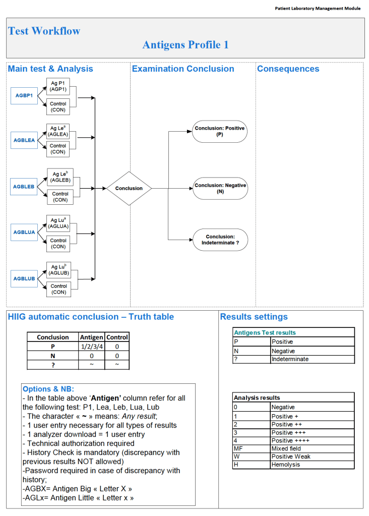
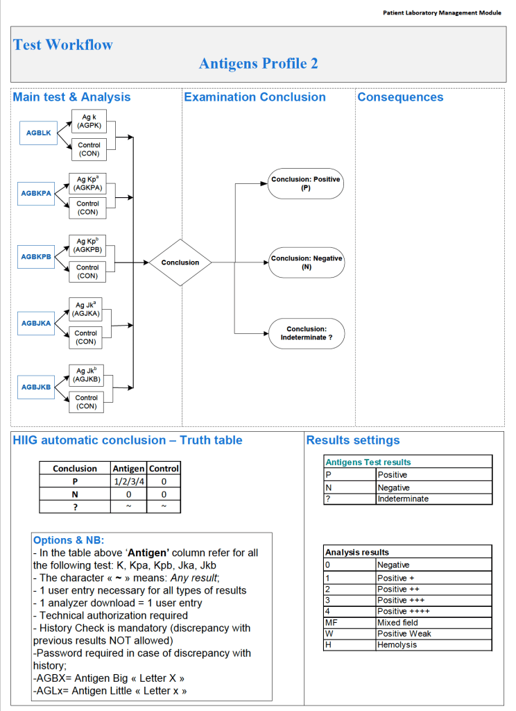
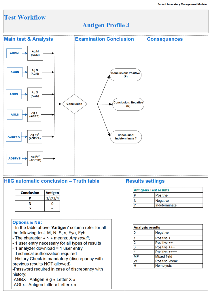
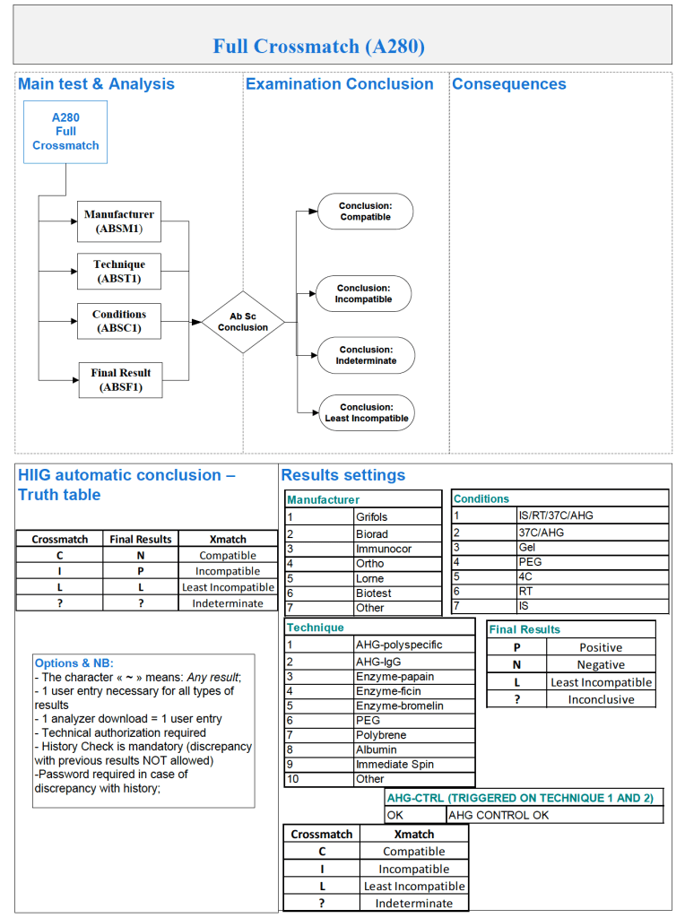
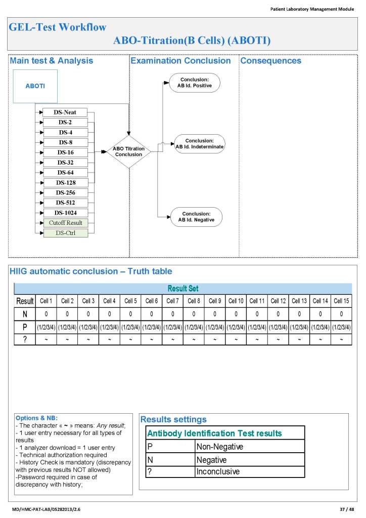
In Medinfo, you store a truth table of possible results and interpretations.  Medinfo will directly read the machine interface data, interpret it according to the rules you make.  The data can be numeric or alphanumeric or both.  Each test, each equipment can have its own unique rules if necessary.

In my opinion, it is best to avoid middleware if your blood bank software can perform this function directly.  When you upgrade it, it is much less likely to show errors—and you only have to deal with one vendor.  Can you be certain that the middleware vendor and the blood bank software vendor will work well together to resolve any issues?

The following are some examples of truth tables, i.e. the rules for interpretation and disposition of interface results actually used at HMC Doha during my tenure there:

Example 1:  Blood Component Production Truth Table

Production can only proceed if the volumes are within the specified ranges.  This is very important if you are going to perform pathogen-inactivation since the ultraviolet illuminator requires a specific range, even more so if you are adding Mirasol (riboflavin) and PAS (platelet additive solution).


Example 2: ABO/D Antigen Typing:

One manufacturer’s requirements:

Example 3:  Complicated D Typing Algorithm:

The acceptable range for automatic interpretation is much more complicated for D typing with the Ortho Vision MAX and uses 3 different monoclonal cocktails:

Example 4:  Direct Antiglobulin Test Algorithm:

Both alphanumeric and numeric results are used.

Example 5:  Four-Cell Antibody Screen:

Truth table for interpretation requires both alphanumeric and numeric results:


Conclusion:

If you are fortunate to have a dedicated blood bank software, you may not need middleware.  Otherwise, you may need to use it for linking to general laboratory systems.  Hopefully, your vendors will cooperate with each other.

To Be Continued:

2/7/20

Processes and Software Building 10: Hospital Blood Bank/Transfusion Services Overview

The following post is based on my experience 2011-2020 at HMC through 16/4/20:

I have had many posts about the Blood Donor Center from registration, collection, processing, testing, and dispatch (inter-depot transfer).  The hospital transfusion service or hospital blood bank continues the process on selection of the appropriate blood component for the patient.  Specifically, it:

  1. Verifies the ABO/D type of RBCs and ABO type of plasma components received
  2. Physically examines each unit checking for leaks, labelling errors, etc.
  3. Receives into stock the various components
  4. Performs basic type and screen (group and save) testing of the patient including ABO/D type and antibody screen
  5. Identifies antibodies if the antibody screen is non-negative
  6. Performs direct antiglobulin test and elution if positive
  7. Modifies components (thawing, aliquoting, irradiating, washing, pooling)—although in some sites, these latter functions may be performed in the Blood Donor Center
  8. Performs compatibility testing and selects the appropriate method (electronic, immediate-spin, antiglobulin phase crossmatch)
  9. Releases blood components to outside staff (nurses, doctors, etc. as allowed by the local authority)
  10. Investigates transfusion reactions

When I was at HMC which included many hospital blood banks, we standardizes our methodologies/processes as much as possible, but we still had some differences based on the equipment at each site.  When we built the blood bank computer system, we had to build a specific process for each test, taking into account the methodology and the type of reagents used.  We used the manufacturer’s recommendations when establishing the criteria for each test.

There were manual and multiple automated tests, e.g. for ABO/D typing.  Rules were established when automated release was allowed and when a manual review was necessary.  Complicated cases were referred to the transfusion medicine physician for review and comment.

In our system, all tests could be ordered and performed from all sites.  Transfusion medicine physicians could review all work from all sites.  For technologists, they were restricted to the sites they worked or supervised except to review results.

All patient results across the entire system from the current and previous system were available and could be used to make/enforce rules.

In general, certain categories of results were referred to the transfusion medicine physician for review, but any test could reviewed by him/her, especially if a clinician requested it.  Everything was documented in the software.

Component modification (thawing, aliquoting, irradiating, pooling, washing) processes were the same at all sites AND the blood donor center.  Each modification changed the ISBT designation of the component, a new ISBT label was printed, and the outdate of the components were updated.

Antibody workups were still performed manually, but direct antiglobulin tests could be  manual or automated.  In each case,  review with an interpretative comment was made by the transfusion medicine physicians and might include recommendations for selection and use of components.  Rules enforced by the software could be made to enforce these recommendations.

To Be Continued:

1/7/20

My Opinion: Use of Enzyme Panels

Working for many years in the Middle East/Gulf, I have encountered significant antibodies that can only be detected at enzyme phase.  This is especially true of Rh system antibodies, particularly anti-c in an R1R1 patient.  I have attached an example.

The reasons I strongly recommend this practice are:

  1. Weak Rh system antibodies (as above)
  2. Confirmation of enzyme-labile antibodies, especially if there may be both enzyme-labile and enzyme-resistant antibodies

It is also important to consider which enzyme to use:  bromelin, ficin, or papain usually and sometimes trypsin or chymotrypsin.  They do not always attack at the same site.

In addition to most common MNSs and Duffy system antibodies, many Kell antibodies (e.g. K or K1, Kpa) are labile with papain but less so or not at all with ficin.

Using enzymes is a double-edged sword since they may enhance cold antibodies and thus cause nonspecific reactions.  Thus, I know many of you may not routinely include them in your workups.

It is essential to follow the manufacturer’s recommendations for their use.  If you make your own enzyme-treated cells and prolong the incubation, you may get false positivity.  You should also be careful about using potentiators with enzyme-treated cells—normally I run them in saline, not LISS!

Since anti-c may cause severe hemolysis and severe hemolytic disease of the newborn, I am especially vigilant in my R1R1 patients, particularly females of child-bearing age and all chronically transfused patients.  I prophylactically match R1R1 patients with R1R1 RBCs in these categories, regardless if either anti-E or anti-c are expressed.

I would be very interested to know your practices?  When do you use enzymes?

1/7/20

Nonspecific Antibodies: My Approach

Anyone reviewing antibody panels, especially in the Middle East/Gulf region, encounters many panels for which no antibody specificity is identified.  As a transfusion medicine physician who often got called during the night for release of RBCs for patients with “nonspecific” pattern, this was a big headache.

Is it “nonspecific” because there isn’t a clinically significant antibody OR the technologist did not perform the testing or its interpretation correctly?  Does it need further testing?  Do I release blood components at this time?

I have attached an example of an antibody panel that has a few weak to 1+ reactions using a gamma-whole-molecule IgG AHG reagent.  Notice that they are enzyme-labile.

I have worked in this region both before and after gel/glass bead technology was introduced.  In the good old days, I formerly only used a gamma heavy-chain specific AHG in tubes with LISS.  Nonspecific reactions were few.

When we adopted gel and at some sites glass bead columns, there was only a choice between polyspecific AHG and whole-molecule IgG AHG.  The rate of non-specificity soared from less than 10% to over 30% of panels.  The reactions were reproducible over multiple technologists at multiple sites.

When I repeated the same specimen against a manual tube panel using gamma-heavy-chain specific AHG, the reactions disappeared in the majority of the cases.  Why?

Heavy chain specific reagents do not detect light chains.  Light chains are the same in IgM and IgG antibodies.  Both polyspecific and whole molecule IgG AHG detect light chains.  Thus, high-thermal-amplitude cold antibodies may be detected with the latter but are unlikely to react with heavy chain specific reagents.

When I used manual tube reagents, I would have missed these reactions, but I do not recall this having clinical consequences except in very rare circumstances (refer to my previous post about the anti-Jka only detectable with polyspecific reagents—I have seen 3 in 30 years).

I have asked various gel/glass bead manufacturers to provide me with heavy-chain gamma-specific AHG, but none have agreed.

Normally, for automation, I use IgG-whole molecule AHG.  If the reactions are nonspecific, we repeat the testing manually using the gamma-heavy-chain-specific AHG.  If negative, I ignore the nonspecific reactions and use the same gamma-heavy-chain-specific AHG for full antiglobulin-phase crossmatching.

In general, with nonspecific reactions, I recommend the following:

  1. Always do enzyme panels, sometimes with both papain and ficin reagents:  many Rh antibodies are optimally detected only at enzyme (example:  R1R1 with apparent anti-E at AHG but the anti-c only seen at enzyme)
  2. Perform an extended Rh/Kell/Duffy/Kidd/Kell/MNSs/P1 phenotype and specifically check for those negative typing results AND for dosage (could the antibody only be detected in homozygous cells like many anti-M are?).
  3. Perform classical room temperature, 37C, and finally AHG phase testing.  Routinely I do not do this since antibodies not detected at 37C are unlikely to be clinically significant.  Sometimes, the AHG phase reactivity is a cold antibody of high-thermal amplitude.
  4. If the Jka or Jkb antigen typings are negative, repeat using a polyspecific AHG reagent.
  5. Use additional panels from multiple manufacturers.  Some reagents detect more nonspecific reactions than others.
  6. Try other potentiators than LISS such as PEG.
  7. Check the outdate of the panel and reagents:  if less than 1 week remaining, consider repeating with fresh reagents and getting a new patient sample.

Finally, if you still cannot define the specificity, consider repeating after several days.  Maybe it is a newly emerging or an anamnestic response.

I emphasize as a physician, I do not care to see all possible antibodies present in the specimen but rather only those likely to be clinically significant.

The Devil is in the Details: Three Antibody Panels for Illustration

As a Transfusion Medicine physician, I reviewed all antibody workups.  Here are three different panels, all appearing to be panreactive,  at AHG phase.  Note the differences:

  1. Autocontrol is positive, enzyme is panreactive and enhanced
  2. Autocontrol is negative, enzyme phase shows no reactions
  3. Autocontrol is negative, enzyme phase is panreactive enhanced

In the middle of the night, if I am called to select blood, I always keep these patterns first and foremost in my mind.

Case 1:

If the autocontrol is positive at about the same strength as the panreactivity.  I can state that this is probably a warm autoimmune pattern WAIHA (although I cannot rule out underlying clinically significant alloantibodies obscured by this pattern and need to do autologous auto-absorption ZZAP).  Both warm autoimmune antibodies and certain drug reactions may cause this pattern.  Use least-incompatible crossmatch matching any significant specificities you found by ZZAP and observe the patient closely throughout transfusion.

Case 2:

If the autocontrol is negative and all reactions are enzyme-labile—and if you live in the Middle East/Gulf region, then you have a presumptive anti-Ge2.  You can safely ignore this antibody and release least-incompatible crossmatch RBCs, regardless of the strength of the reactions.

Case 3:

If the autocontrol is negative and the reactions are unchanged or enhanced by enzyme, BE AFRAID, VERY AFRAID!!  This is an antibody to a high-incidence antigen.  These can be very dangerous.  In the Middle East/Gulf region, consider anti-H, anti-k (cellano), anti-Kpb, anti-PP1Pk, and rare antibodies to the MNSs such as anti-U or other MN system deletions.  There are many other possibilities, e.g. anti-Fy3.

You need to perform extended antigen typing across the major Rh antigen, Kell, Duffy, Kidd, MNSs, P systems (at least P1):

  1. Run H lectin to rule out Bombay Oh or Parabombay.
  2. Some Rh system deletions and Rh null show pan-Rh reactivity—check D, C, c, E, e typings.
  3. Anti-k (cellano) will be suggested by k-negative phenotype.
  4. Unusual, weak or absent reactions with M and N reagents suggest something like En(a)-negative or similar.
  5. Absent P1 with no other findings, you must rule out anti-Tja (anti-PP1Pk).

I have seen all of these specificities during my time in the Middle East.  All of these antibodies can be clinically significant and often life-threatening.

Conclusion:  NEVER NEGLECT TO REVIEW THE AUTOCONTROL!!!

Nonspecific Reactions, Reagents Near Expiration

Whenever I had a “nonspecific” antibody, I had to first rule out issues with the reagents themselves. The following example shows weak to 2+ reactions in the panel cells and autocontrol.

The variability in the reactions made me initially uncomfortable about called this WAIHA. I then checked the panel details: the testing was done only six days before the panel outdate.

I told my staff to repeat the workup with the new panel expiring five weeks later. The difference is astounding!!

Remember: if you work in the Middle East, the environmental conditions can be extreme in summer (>50C). Do you know how your reagents were handling during transport?

My advice: if you are concerned there is a clinically significant antibody but cannot discern it, consider repeating the workup using fresh reagents.