Blocking Antibodies

This is a revised version of a previous post.

If there is strong antibody binding to an RBC, this may interfere with a typing reagent attaching to the cell and cause a false-negative, i.e. a “blocking” antibody.  Such cells may interfere with the indirect antiglobulin test IAT, i.e. the antibody screen.  The autocontrol and direct antiglobulin test DAT will be strongly positive.

The manufacturer’s instructions for using its reagents should be strictly followed in the presence of a strongly positive DAT.  If there is no reaction with the typing reagent, the result must be indeterminate.

One could try a (relatively) nondestructive elution method such as gentle-heat elution to remove some of the antibody and then retype the cells.  I have found this to be a simple and effective method for my staff to use.  Just remember that despite being “gentle,” there will still be some hemolysis present, but here it is the cells we are trying to save.

Usually, we find this situation in a neonate born of a mother with anti-D.  The baby has a strong DAT but the D typing is negative.  Check the D control carefully:  if it is positive, the result is indeterminate, try another method.  Usually gel/glass bead methods are subject to less interference.  Finally, there is always the classic saline anti-D!

In Medinfo software with a blocking antibody, a nonnegative control will trigger a manual review of the results.  There will be no automatic release.

Here is my process for handling blocking antibodies, which I set up for HMC Doha:

INTERIM POLICY:  ANTIGEN TYPINGS IN PRESENCE OF STRONGLY POSITIVE DIRECT ANTIGLOBULIN TEST (DAT):  RULE OUT BLOCKING ANTIBODY

Principle:

Antigen typing of cells with large amounts of coating antibody (i.e. strongly positive DAT 3-4+) may not always be possible because the bound antibody may block available antigen sites.  This policy is to clarify how to recognize and handle such situations.

Policy:

  1. Always follow the manufacturer’s instructions for the use of the typing reagent.
    1. In particular, note whether a control must be run with the test (e.g. D-control, D-diluent, etc.) or if it is included in the gel or glass bead card.
      1. If a control is required, use exactly what the manufacturer recommends.
      2. DO NOT SUBSTITUTE ANYTHING ELSE AS THE CONTROL!!
  2. Interpret the reactions exactly as the manufacturer indicates.
  3. If the test is invalid because of the control or any other reason, report the antigen typing as indeterminate and send for Transfusion Medicine Physician TMP review.
  4. If the DAT is 3-4+ and the antigen typing shows no reaction (apparent negative), send the case to the Transfusion Medicine Physician for review and final interpretation.  DO NOT ENTER THE RESULT AS NEGATIVE UNLESS THE TMP INSTRUCTS YOU TO DO THIS!!
  5. To rule out a blocking antibody, a special elution to gently remove the coating antibody may be needed so that the RBCs can then be typed (not acid glycine technique—rather use gentle heat elution.)  The Transfusion Medicine Physician will decide whether to do this additional testing.

References:

  1. Standards for Blood Banks and Transfusion Services, Current Edition, AABB, Bethesda, MD, USA
  2. Technical Manual, Current Edition, AABB, Bethesda, MD, USA
  3. Guidelines to the Preparation, Use, and Quality Assurance of Blood Components, European Committee (Partial Agreement) on Blood Transfusion (CD-P-TS), Current Edition
Overextending Oneself—Two Case Studies in the Blood Bank

One has to learn when enough is enough.  There are times when there are staff shortages but the conscientious staff wants to be the Super-Tech and handle all the work, whether or not there are sufficient resources.  This is a big gamble, and there may be serious consequences for the over-achiever and for the patient.

Anecdote #1:  Chicago Blizzard of 1979 (13-14 January):

When I was in my residency training in Chicago, I was in the blood bank during the blizzard of January, 1979.  The following tragedy occurred.

Suse was one of the best blood bank technologists that I have ever known, extremely conscientious and very meticulous—and very fast at doing things.  She was a workaholic.  Suse’s whole life centered on her job at our academic medical center—so much so that she had an apartment near the hospital complex.  In mid-January, a snow storm was predicted with an estimated snowfall of about 5 cm. total.  Actually, that night a blizzard developed and around a meter of snow fell with white-out conditions and zero visibility.  Preoperative patients had been admitted the night before based on the low snowfall prediction.

The next day was chaos.  Essentially only staff who lived near the hospital complex could report to work.  Suse came in and saw all the pending preoperative blood requests.  She decided to “double-up” and work on two cases at one time.  In the rush, she mixed up test tubes and issued ABO-incompatible blood for a surgical case.  The surgeon noted the abnormal oozing of blood at the operative site and stopped the transfusion.  Hematuria developed, but the patient survived.

Suse was suspended pending investigation.  Based on her excellent work record, she was offered to return to work.  Unfortunately, she became very depressed and was afraid to return since she feared she would make another mistake.  She never worked in the blood bank again.

Anecdote #2:  Shortage of Blood at Major Hospital:

1991 in another country, a large hospital complex was suffering a shortage of blood.  A large number of donors were called and the available staff were overwhelmed with work.  One donor phlebotomist decided to collect whole blood from two different donors simultaneously and in the confusion, mixed up the sample tubes for donor marker testing.

Unfortunately, one of those donors was HBsAg positive, but with the specimen mix-up was marked as negative.  The unit of blood was transfused, and the recipient developed fulminant hepatitis B and died.

Analysis:

In both these systems, there were processes in effect not to work on two patient specimens or collect two donors at one time, but the staff took short-cuts.

No one is super-human.  Don’t try to cut corners and handle more than one patient at a time.  Your intention may be good, but you will be judged by the consequences.  No one will care about the extenuating circumstances.  You will be blamed.  I tell my staff that if they cannot handle the workload, they should contact me as the Division Head, Transfusion Medicine, to triage the cases for them.  My role is to bring these events to the higher authorities to get the resources we need to do the work properly and safely.

Therapeutic Apheresis Process

This is an update of a previous post.

Principle:

All therapeutic apheresis procedures are potentially life-threatening and must only occur by an order from a transfusion medicine physician with experience/competence in such procedures.

Definitions:

  • Referring Physician is the clinical physician requesting a therapeutic apheresis procedure.
  • Transfusion Medicine Physician is a physician in the Transfusion Medicine Section with medical privileges for therapeutic apheresis procedures.  This includes the Head, Transfusion Medicine, consultants in Transfusion Medicine, and designated specialist physicians in Transfusion Medicine.  The final decision to accept/reject the patient is made by the transfusion medicine physician.
  • Covering Physician is the clinical physician designated by the referring physician to be physically present and covering the patient in case of any adverse reactions during a therapeutic apheresis procedure.
  • Apheresis Nurses are nurses in Transfusion Medicine who are designated by this section for performing therapeutic apheresis procedures.
  • Medical Privileges are determined by the Department of Pathology and Laboratory Medicine in conjunction with the medical staff medical privileging by the Hospital Medical Director.

Policy:

  1. The referral physician will discuss the request for a therapeutic apheresis with the designated transfusion medicine physician.  The referral physician must certify that the patient can tolerate the procedure based on his medical condition.
  2. The transfusion medicine physician will review the patient’s clinical and laboratory data, with special note of the history of allergies, medications, previous transfusion reactions, and current vital signs.
  3. Vascular access will be initially assessed by the apheresis nurse.  Any questionable situations will be reviewed by the transfusion medicine physician.
    1. It is the responsibility of the referring physician to provide vascular access.
  4. The following laboratory values (less than 24 hours old) must be available before the procedure may begin—additional tests also as per transfusion medicine physician:
    1. CBC including platelet count
    2. PT and APTT
    3. Fibrinogen
    4. Serum calcium
    5. Serum protein and albumin
    6. LDH for TTP cases
  5. A valid type and screen must have been done within the previous three (3) days of the procedure.
  6. Upon review of # 2 through 5, the transfusion medicine physician will determine if the procedure is indicated and will communicate this to the referral physician, who will sign written order in the patient chart.  Appropriate replacement fluids will also be mutually agreed upon in advance of the procedure and ordered by the transfusion medicine physician.  The order specification must include:
    1. Name of procedure and specification (e.g. therapeutic plasma exchange, isovolemic)
    2. Replacement fluid type and volume (e.g. 3 liters 5% albumin, 2 liters, FFP, cryoprecipitate, normal saline)
    3. Blood component orders if indicated (e.g. RBC exchange) and timing (before, during, and/or after the procedure)
    4. Calcium replacement (e.g. 2 grams calcium gluconate IV in 100 ml normal saline to run during the procedure)
    5. Any special laboratory testing post-procedure
  7. Use of a remote vital signs/oximeter/EKG device such as the Umana T1 should be considered in high-risk patients.
  8. The apheresis nurse takes orders ONLY from the responsible transfusion medicine physician.
    1. If the referring physician wants any other orders or changes, he must get the approval of the responsible transfusion medicine physician.  He does NOT give orders to the apheresis nurse
  9. The apheresis nurse will follow the orders of the necessary prescribed replacement fluids (FFP, albumin, PPF) in the quantities necessary for the exchange..
  10. The referring physician (not the apheresis nurse) will obtain the signed, informed consent from the patient.
  11. If vascular access is unsatisfactory, the referring physician will obtain the proper access (central line, AV shunt, etc.).
  12. The referring physician will arrange for a physician member of his team to be present at the actual therapeutic procedure.  This physician designate will be responsible to treat any complications arising from the procedure.
  13. Vital signs and weight will be obtained before starting the procedure.
  14. When approved by the Blood Bank Director or designate with proper venous access and informed consent, the apheresis may start the procedure in the presence of the patient’s covering physician.  The procedure will be performed in a designated hospital area.
  15. The procedure must be documented on the appropriate therapeutic apheresis order and procedure worksheets.

References:

  1. Standards for Blood Banks and Transfusion Services Current Edition, AABB, Bethesda, MD, USA
  2. CAP Standard TRM.42245 regarding therapeutic apheresis procedures

Processes and Software Building—Part Two

This is a revision of a previous post.

Documenting Processes:

My previous post emphasized how important it is to map the current state across all processes as the first step to optimize current operations and prepare for a new computer system.

One non-blood bank vendor submitted the following as a complete representation of all current processes—across more than 4,000 tests and hundreds of instruments:

  1. Order something
  2. Receive specimen
  3. Perform test
  4. Report test

This was the same for each of the tests in the different sections of the laboratory—be it blood bank, anatomic pathology, chemistry, hematology, etc.  I was flabbergasted!  What were we paying for?

As Head of the Laboratory Information Systems, I rejected this.  I would have been ashamed to submit this to a client as a sufficient current state.  Even more astounding was the fact that that vendor actually mainly used the same four-step flow chart for the tests in their new computer build!!

As painful and time-consuming as it is, one must develop a specific flow for each process.  This could include:

  1. Specimen condition and acceptability criteria
  2. Possible results for each part of the test
  3. Interpretation of each result
  4. Control results
  5. Acceptability criteria
  6. Truth table
  7. Reflex testing triggered by the results

When we built our first dedicated blood bank computer system, the company would take a module and completely map out the current processes collaboratively with me.  After this, I analyzed the critical control points and started to map out the improved computer processes that would take over.  I did not want to throw out the successful manual system, just to optimize it.  After that we would build that those limited processes in the software and test it.  If it failed, we would correct it, and the vendor didn’t charge us extra for the corrections.  It was a beautiful collaboration.

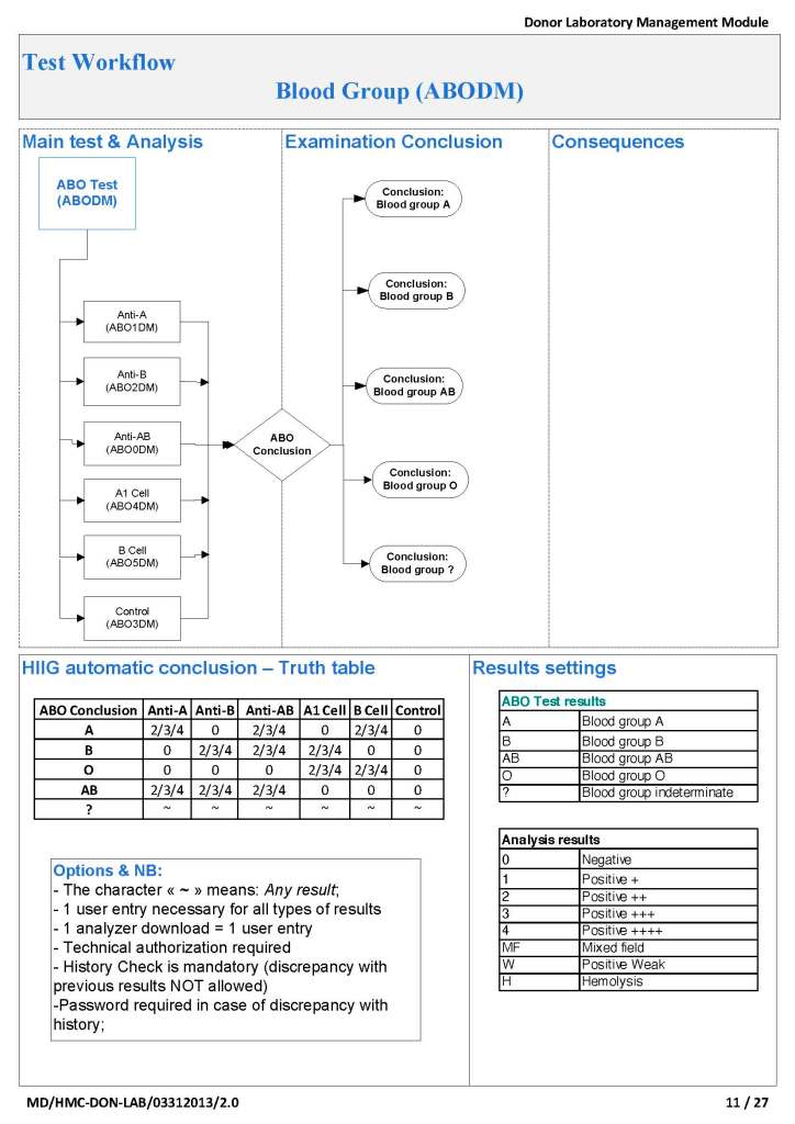
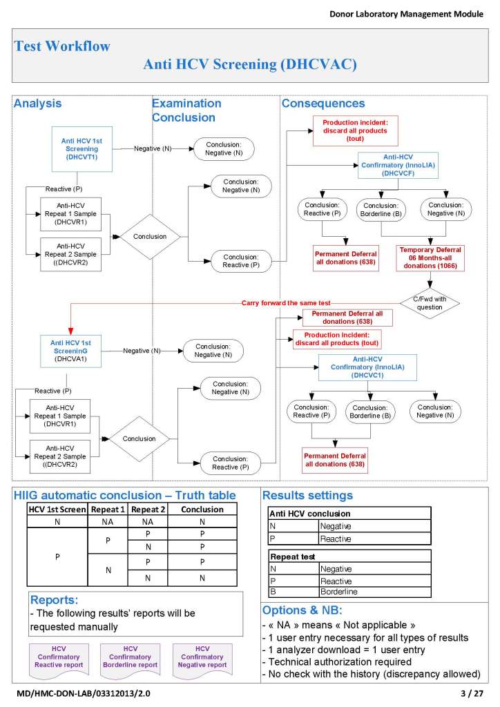
To illustrate these points, I am showing two process flows from our Medinfo Hematos IIG build:  one for the ABO typing (forward and reverse) for donors and the other a complex testing algorithm flow for HCV donor marker testing.  These are from previous builds and have been updated subsequently.

ABO Typing: Attachment One

This consisted of six individual tests forward (anti-A, anti-B, anti-A,B), two reverse (A1 cells and B cells) and a control.  The acceptable tests for automatic typing were in {0, 2, 3, 4}, other results (mixed field, weak, 1+, hemolysis) required a manual interpretation.  There is a truth table for interpretation of all six results together.

Donor HCV Testing:  Attachment Two

This is a more complicated flow that includes multiple tests (HCV-antibody EIA, HCV-LIA, and HCV-NAT).  Results may trigger reflex testing immediately (abnormal HCV EIA triggers HCV-LIA, abnormal HCV-NAT triggers HCV-LIA, etc.) or repeat testing after six months for indeterminate results.

In each case, every possible result is listed and its interpretation and acceptability criteria.

In summary, it may take considerable time to map out all your processes, but this is time well spent and allows you build your system accurately.  There will be few surprises this way.

Case Report: Overwashing During Elution

Note:  This is a repost.

I cannot emphasize enough proper technique in doing the washing during the elution process.  We are usually concerned about too little washing and thus possibly residual reactions in the last wash.  However, aggressive overwashing may remove the bound antibody resulting in a negative result.

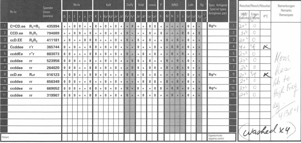
Here is an example of anti-PP1Pk (alias anti-Tja).  The mother’s panel shows an antibody to a high prevalence/incidence antigen with negative autocontrol and no lability at enzyme phase:

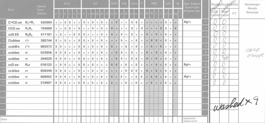
The neonate’s DAT was weak positive at polyspecific and IgG monospecific phases.  An eluate was performed.  Here is the result after washing four (4) times:

Since 2 cells in the last wash were very weakly positive, the washing was continued for a total of 9 times with the following results:

Even then there was very weak positivity in one cell, but the eluate was negative.  We had washed away the attached antibodies.

A Novel Way to Document Therapeutic Phlebotomies in the Blood Bank Computer System

Therapeutic phlebotomies TP, like regular whole blood donation WBD, both require collection of whole blood into a blood bag set.  In whole blood donation, we ask two questions during the process:

  • Is it safe for the donor to donate?
  • Is it safe for the collected unit or its processed components to be given to recipients?

For TP, we only have to consider the first question so the process must ensure the patient/donor’s safety.  We do not have to concern ourselves with the use of the collected product—it will be discarded.

Since the donor collection processes of both TP and WBD are similar, why couldn’t we use the blood bank computer software to document the TP procedures?  The process is a subset of normal WB donation.  On this basis, I make suggestions on using the donor module to document the TP process.  It is basically a truncated version of blood donation process in the Medinfo Hematos IIG system:

  • Registration
  • Donor Safety
  • Vital Signs
  • Hemoglobin Determination
  • Blood Collection Data
  • Adverse Effect Reporting
  • Discard of Unit
  • Documentation of Physician’s Order and Transfusion Medicine Physicians Acceptance

Registration:  Positive patient identification can be made through the donor registration process;  force selection of an inexpensive bag type (not the Reveos set) for this purpose.

Donor Safety:  Perform a modified, shortened donor questionnaire covering the medical history and medications is used.  Confirm that the patient has had food and drink before donating.  Require a waiting period of 24 hours before the next procedure.

Vital Signs and Weight:  Measure weight plus BP, pulse, temperature, and respiratory rate as well as inspect the arm for scarring before procedure.  Allow repeat vital signs monitoring after the procedure if requested by the transfusion medicine physician.

Hemoglobin Determination:  Allow acceptable Hgb >= 11 g/dl or >33% hematocrit

Blood Collection:  Use the same process for the mixer-shakers but the amount collected can range up to 500 ml with amounts <405 ml acceptable for small patients

Adverse Effect Reporting:  The complications of TP collection are the same as WBD.  Use the same system as for WBD.

Discard of the Unit:  Print discard label and quarantine of the ISBT unit number in system (so that it cannot be used for transfusion).

Documentation of Order:  Create separate fields for the ordering physician and for the approving transfusion medicine physician.  Capture scan of paper orders and incorporate into the TP computer encounter.

Other Considerations:  In high-risk cases, e.g. with pre-existing cardiovascular, pulmonary, or cerebrovascular disease, one could consider using a remote monitoring device such as the Umana T1 device to record vital signs, EKG, and oxygen saturation that can continuously record these parameters and trigger user-definable alarms during the process and afterwards if desired.  The data can be incorporated into the blood bank computer encounter.

Antibody Titration

My practice across the globe has exposed me different rationales to performing antibody titration.  In my American training and practice (and also at international institutions following the American version of AABB accreditation), I only routinely performed titration of anti-D for Rh(D) hemolytic disease of the newborn and anti-A/anti-B for ABO-incompatible stem cell transplants AND ABO-incompatible renal transplants.

I have had heated arguments with some physicians who insisted they wanted titers for other antibodies.  The AABB Standards do not require this but leave it to the discretion of the Transfusion Service Medical Director.

In my entire career, I never worked in a blood bank or blood center which had optimal staffing or resources.  I focused on what was medically/technically necessary and even then still had shortages.  If performing a test does not change the clinical treatment, why perform it unless you are doing a research project!

Titration is a time-consuming, and until recently, a tedious manual task.  Recently some of the automated immunohematology analyzers offer a titration program.  We used the Ortho Vision Max which could perform both IgG and IgM titers within one hour—walk away!!  However, during that time, the titration procedure monopolized the analyzer.

Nowadays, low-anti-B-titer group A universal plasma and low-titer (anti-A and anti-B) group O whole blood may be offered as components.  At HMC Qatar, a preliminary study showed about 50% of units could be classified as low-titer (defined as a saline titer <1:128).  The amount of titration will require an automated analyzer.

The ABO-incompatible renal transplant program at HMC Qatar was modelled after Sweden’s Karolinska Institute.  However the latter site performed manual IgG and IgM titrations using Biorad/Diamed gels.

I did not have sufficient resources to commit staff to manual titration at HMC so I did a comparison study between the Ortho Max and the Biorad manual gel methods.  We were able to get good correlation and used the automated method for the transplant.

I still don not perform against performing titrations for antibodies other than anti-D.  I always ask, ‘Does the titration correlate with clinical severity?’  Unlike anti-D, antibodies such as anti-Kell and anti-c may be low titer but cause death.  Can anyone show me a definitive study that titers are useful except for transplants and Rh(D) hemolytic disease of the fetus/newborn?

Since the method was working well on the Ortho equipment, I next established an interface to Medinfo.  The test was performed separately for IgG and IgM antibodies.  Medinfo recorded the reactions in all the wells.  The last well showing a 1+ reaction was interpreted as the titer (e.g. if 1:64 were the last 1+ reaction, then the titer was 64 in Medinfo).

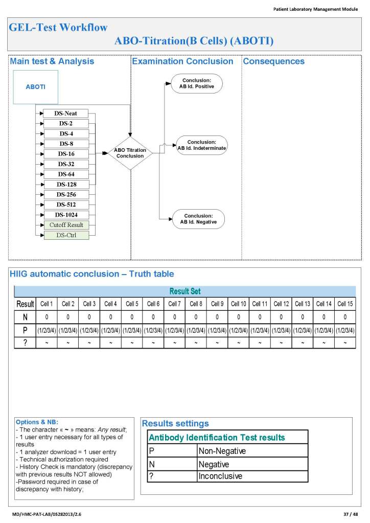
The Medinfo process is shown below.

My Opinion: Use of Enzyme Panels

This is an updated version of a previous post.

Working for many years in the Middle East/Gulf, I have encountered significant antibodies that can only be detected at enzyme phase.  This is especially true of Rh system antibodies, particularly anti-c in an R1R1 patient.  I have attached an example.

The reasons I strongly recommend this practice are:

  1. Weak Rh system antibodies (as above)
  2. Confirmation of enzyme-labile antibodies, especially if there may be combinations of enzyme-labile and enzyme-resistant antibodies (e.g. anti-Fya and anti-c).

It is also important to consider which enzyme to use:  bromelin, ficin, or papain usually and sometimes trypsin or chymotrypsin.  They do not always attack at the same site.

In addition to most common MNSs and Duffy system antibodies, many Kell antibodies (e.g. K or K1, Kpa) are labile with papain:  however, with ficin they may be partially labile, unaffected, or even enhanced.

Using enzymes is a double-edged sword since they may enhance cold antibodies and thus cause nonspecific reactions.  Thus, I know many of you may not routinely include them in your workups.

It is essential to follow the manufacturer’s recommendations for their use.  If you make your own enzyme-treated cells and prolong the incubation, you may get false positivity.  You should also be careful about using potentiators with enzyme-treated cells—normally I run them in saline.

Since anti-c may cause severe hemolysis and severe hemolytic disease of the newborn, I am especially vigilant in my R1R1 patients, particularly females of child-bearing age and all chronically transfused patients.  I prophylactically match R1R1 patients with R1R1 RBCs in these categories, regardless if either anti-E or anti-c are expressed.  I have seen many examples where the anti-c is only detected at enzyme phase.

It is my practice to always include an enzyme panel.  I would be very interested to know your practices?  When do you use enzymes?

My Opinion: Issues in Transfusion Medicine Software and Component Production

I anticipate that there are several innovations coming or in the process of coming to mainstream blood component production and software.  Some of these I have already addressed in some of my previous posts:

Pathogen inactivation:  We have had this for over a decade.  However, with new emerging pathogens, this will become more important so I expect it will be adopted in many centers where it is not currently being used.  I expect we will close the loop and pathogen-inactivated RBCs will be available so all components will be treated.  Still, the first-generation pathogen-inactivated RBCs may have reduced shelf life compared to regular, untreated units.

Automated component production:  Although this is expensive, it does provide excellent GMP production.  It is fast and may provide higher yields, especially for platelets.  I expect more centers will adopt this technology, especially in combination with pathogen inactivation.

Blood bank computer software:  This software must be considered as dynamically changing, and considerable resources are needed to keep in compliance with ever-changing international regulations and the latest epidemiologic data.  Production rules can be strictly and mercilessly enforced by a dedicated blood bank computer software.  It can also ensure that the final ISBT label is not applied unless all the production rules (registration, collection, processing, and testing) are met.  Manual processing is extremely risky nowadays with all the parameters to be monitored.

Patient Blood Management:  Current blood bank software does not adequately address the need for prospective review of component orders.  I expect that collaboration will occur between laboratory and blood bank software vendors to fill this gap.

Refrigerated platelets:  The pendulum swings back to this component which was used over 40 years ago.  Refrigerated platelets suspended in additive solution may be effective up to 14 days for hemostasis in the trauma setting.  These platelets are activated so standard 20-24C stored platelets may be preferred for prophylactic transfusions.

Low-titer group A universal plasma:  This is already available, but its use will increase because of the low numbers of group AB units available and increased demand.  This includes its production for COVID convalescent plasma.  Your transfusion medical director must decide what “low titer” means.  Also you need a robust way of performing anti-B titers, this may require use of an immunohematology analyzer with titration built-in.

Low titer group O whole blood:  Use of this product may reduce the need for components in massive transfusion settings but it requires performing anti-A and anti-B titers on large numbers of units.  Your transfusion medical director must decide what “low titer” means.  Also you need a robust way of performing anti-A and anti-B titers, this may require use of an immunohematology analyzer with titration built-in.  Also, you must decide whether to leukodeplete the whole blood units:  few whole blood filters are platelet-sparing.