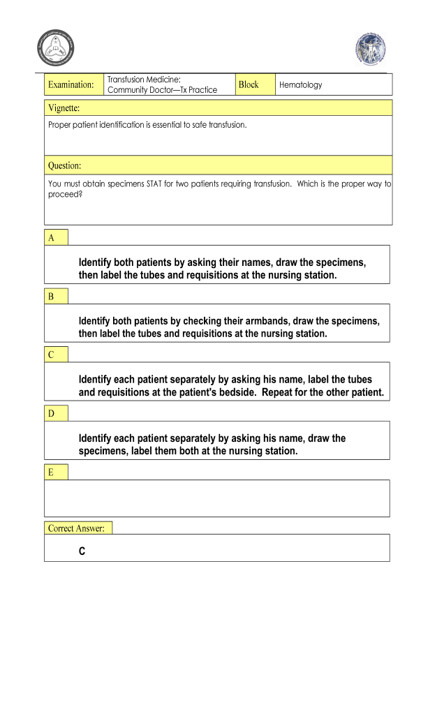
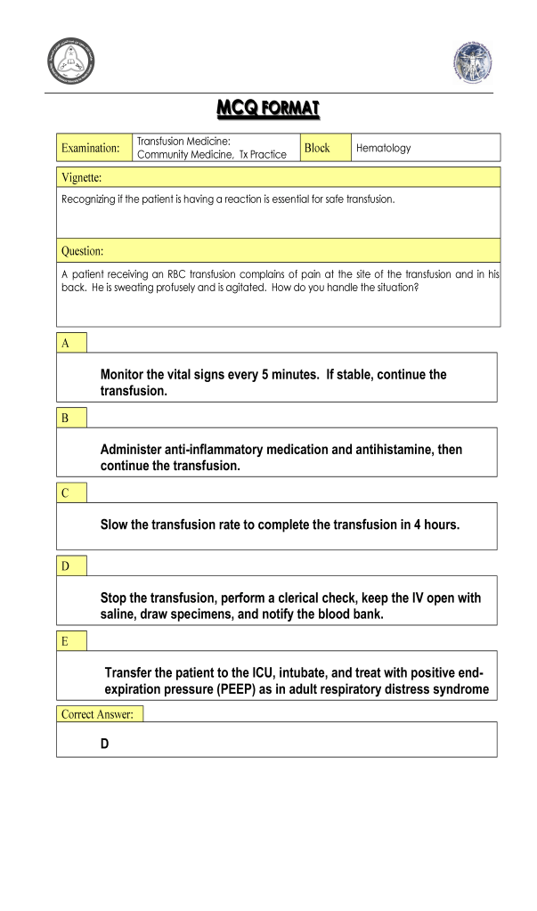
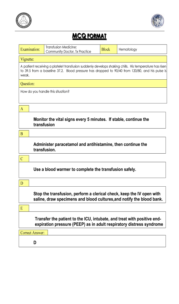
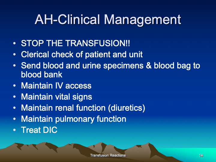
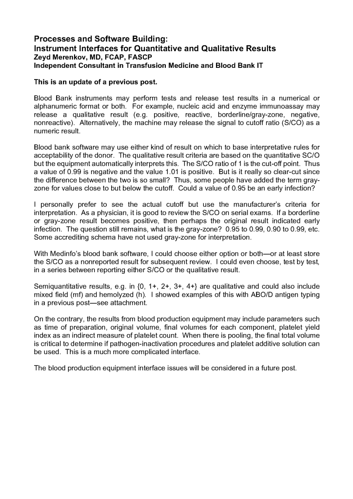
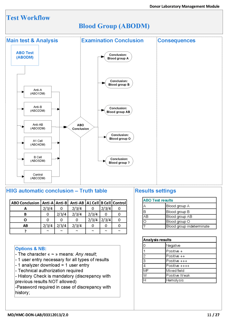
This is the first part of a medical student lecture I gave at National Guard Health Affairs in Riyadh. The lecture was given before we had a blood bank computer system and shows manual processes.


Includes compatibility testing (AHG, electronic, immediate-spin), antigen typing, antibody screening and identification, direct antiglobulin testing, elution, transfusion reaction and drug reaction workups, component typing tests upon receipt in hospital blood bank, release and return of components
This is the first part of a medical student lecture I gave at National Guard Health Affairs in Riyadh. The lecture was given before we had a blood bank computer system and shows manual processes.


Rh immunoprophylaxis has big effect on the Transfusion Medicine operations. With a very active obstetrics program, there are numerous prenatal blood bank workups and administrations of Rh immune globulin RhIG—and consequently many antibody identifications. In fact at my previous position, the largest single source of antibody workups was from patients post-RhIG administration.
As a result of the antenatal RhIG use, there were numerous positive antibody screens. Our policy had been to always do a full antibody identification, both AHG and enzyme panels, and this made up the bulk of our antibody testing. In my experience, I have found other antibody specificities in some of these patients, including anti-Kell and anti-c, both of which could be very clinically significant.
As part of the workup, we also did ABO and extended Rh and Kell phenotyping, and thus identified rare Rh phenotypes (r’r’, -D-, etc.) and several Bombay phenotypes.
Unfortunately, I have been at institutions where they assumed a positive antibody screen in a patient with recent RhIG administration was passive anti-D and did NOT perform further testing.
If the patient needs blood and the antibody screen is positive, the antibody workup must be performed for routine release. This means about a 20-30 minutes delay in release until the antibody panels are completed and reviewed. Otherwise, blood can be released through an emergency protocol if the clinician accepts responsibility for incomplete testing at the time of release.
Many clinicians refused to do this emergency release and insisted on waiting for the antibody identification before accepting RBC components. They would not take the responsibility for the emergency release.
If we had done the antibody workup in these RhIG patients, we would have identified the anti-D, and I as the transfusion medicine physician would then advise the clinician to accept emergency release of RBCs while we complete the antibody workup.
For this reason alone, I insisted on a full antibody workup for all RhIG patients RhIG who exhibit a positive antibody screen. I would be comfortable in recommending emergency release if I had results of the previous antibody identification showing passive anti-D. The delay in release could adversely affect the patient’s outcome if there is active bleeding.
In summary, if a patient has a positive antibody screen post RhIG administration, you should still do ABID to rule out other antibodies and facilitate release of RBCs to a bleeding patient.
As a transfusion medicine physician, I must know if I can trust my staff’s interpretation of immunohematology testing. I may be called at night and they will provide me with results and I must use these to make a medical judgment. If their interpretation is flawed, I might make a decision that harms the patient.
I really don’t like multiple-choice questions, but nowadays this is often the norm. For my staff, especially senior staff and those who want to be promoted to senior staff, I have developed a series of projective exercises to help me understand their thought processes. I also used this approach on prospective candidates for hire.
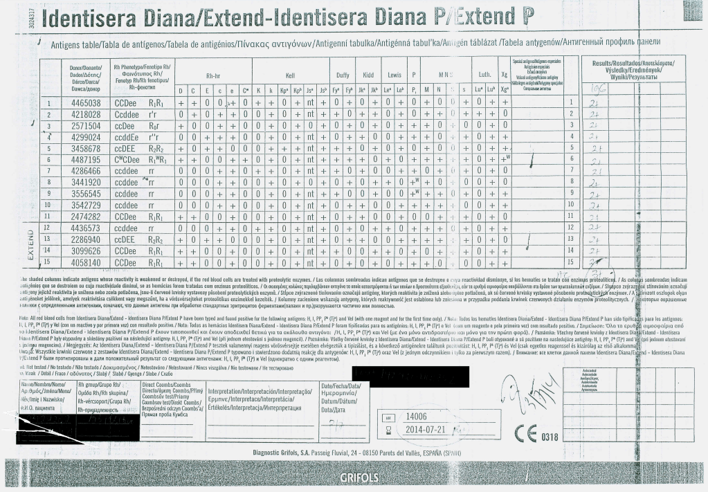
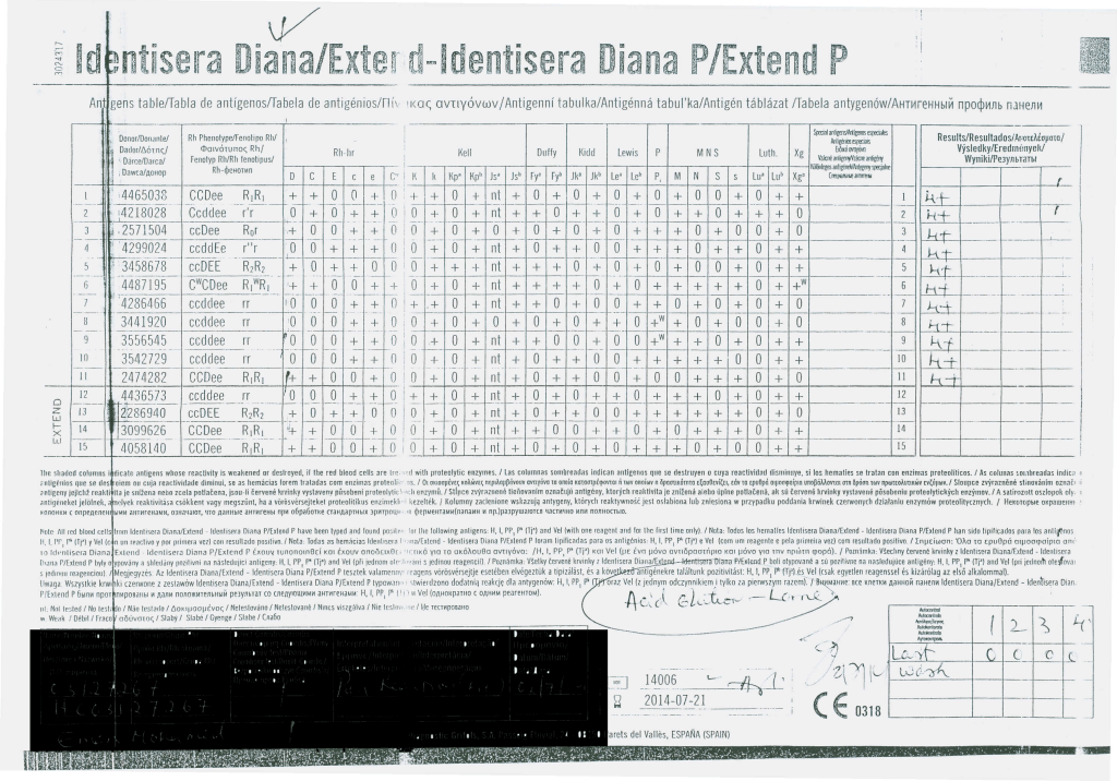
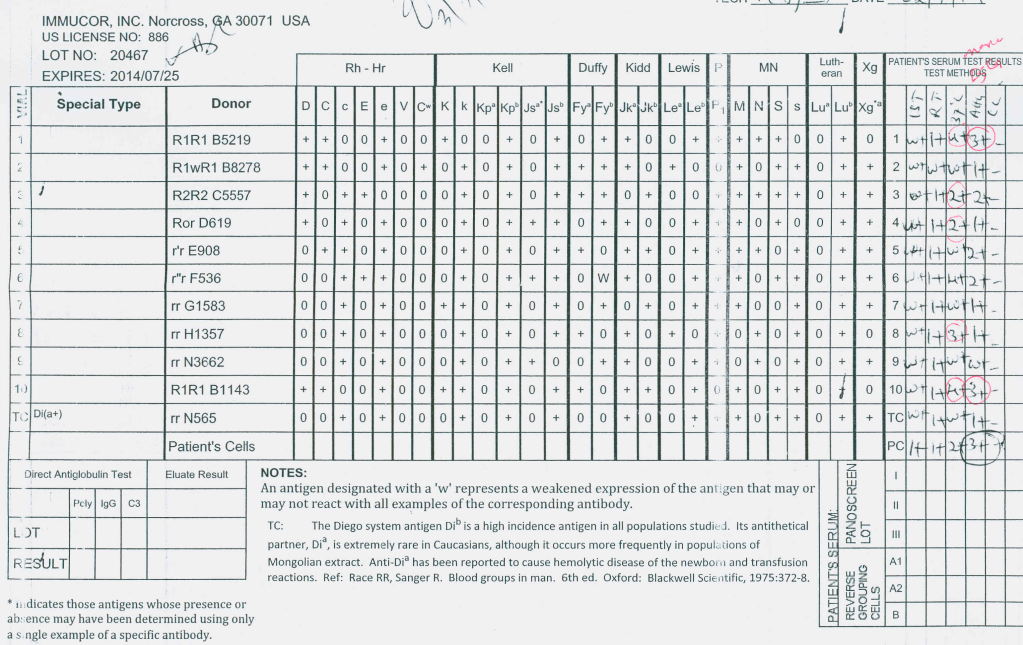
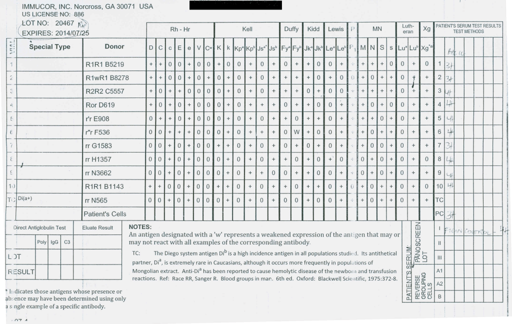
Here is a sample exercise. I have the candidate or staff review this panel and tell me to interpret it:

Most of them answer that this is an anti-Cw without hesitation. However, they are basing that on only one Cw-positive cell.
More astute ones indicate it might be anti-Cw but ask to test additional Cw-positive cells and perform an enzyme panel. These are the ones that I will consider for hire or promotion now.
This is the start of a series of posts on how Medinfo blood bank software was designed for product delivery in the HMC system in Doha.
The overall process was:
It was also possible to release blood components directly from the Blood Donor Center to HMC hospital blood banks as a contingency. Client hospitals outside the HMC system still had to obtain their components from HGH General Delivery Deposit.

Antiglobulin reagents are used to detect molecules bound to the RBC surface. What they detect depends on their specificity. Such detection can be performed in the routine immunohematology laboratory or elsewhere such as flow cytometry. This discussion is for the blood bank laboratories, both routine and reference immunohematology.
This is how I classify and use the various reagents in my daily practice:
There are many types of antiglobulin reagents:
Whole molecule IgG detects class-specific mu heavy chains AND light chains kappa and lambda. Since kappa and lambda are found on all immunoglobulin classes, whole molecule reagents can detect IgM so there may be weak staining with cold antibodies that are not clinically significant.
C3d is the final breakdown product of C3b and does not cause hemolysis. Its presence merely means that at some time—unspecified—complement was fixed. C3c is an intermediate product in the breakdown pathway. If detected, C3c positivity means ACTIVE complement fixation was occurring at the time of specimen collection.
General Use:
Routine DAT testing:
Antibody workups:
Routine: antibody screens and AHG crossmatch (if indicated)
Monospecific gamma heavy chain is preferred to minimize non-clinically significant, cold antibody interference.
Complicated—where detection of complement reactivity is especially important:
Polyspecific for drug-related hemolysis and transfusion reactions
Difficult antibody workups, e.g. to rule out anti-Jka and/or anti-Jkb
Specialty Reference Procedures:
Complement fixation may be important in various drug-related hemolysis and some transfusion reactions so I always use a polyspecific reagent in these situations. Most antibodies can be detected by gamma heavy-chain specific reagents; however, there are rare examples of anti-Jka and anti-Jkb which are only detected by complement. Whenever I have a nonspecific reaction in an Jka-negative or Jkb-negative patient, I repeat the AHG panel using polyspecific reagents. I do not use polyspecific routinely because of the nonspecific and non-clinically significant cold antibodies.
One way to assess for the clinical significance of an antibody is to determine its IgG subclass. In general, IgG3 antibodies may fix complement and cause severe hemolysis. Both IgG1 and IgG3 antibodies cross the placenta and may cause hemolytic disease of the fetus/newborn.
In summary, when reviewing immunohematologic reactions using AHG, I always remember to check which type of AHG reagent was used. I always keep multiple types of AHG reagents in the laboratory for the reasons explained above.
This is a revision of a previous post, incorporating an updated definition of Transfusion-Associated Cardiac Overload TACO from the National Healthcare Safety Network, Biovigilance Component Hemovigilance Module Surveillance Protocol, Â Version 2.6.


This is part 3 of a medical student lecture I gave at National Guard Health Affairs.


This is the second part of a lecture I gave to medical students at National Guard Health Affairs in Riyadh.


This is an updated version of a medical student lecture I gave at National Guard Health Affairs in Riyadh.


To be continued
I am a strong believer in performing both AHG and enzyme panels together in routine antibody workups. I especially feel this is important when the patient is R1R1 since I always want to rule out anti-c. Sometimes, anti-c is only identified in the enzyme phase.
This is a case from my files of an R1R1 patient with the following results:
Antibody Screen:

AHG Panel:

Enzyme Panel (Ficin):

This anti-S is enzyme-labile but anti-c is revealed, only reacting at enzyme phase. The patient was Kell-negative so I selected S-negative, R1R1 K-negative RBCs for transfusion. Anti-c can be a dangerous antibody causing severe hemolytic disease of the fetus/newborn and sever hemolytic transfusion reaction. If only the AHG panel had been performed, the anti-c would have been missed.