Therapeutic Phlebotomy Process–Updated

Principle:

Therapeutic phlebotomy is a medical procedure that requires a written physician’s order and review/approval by a transfusion medicine physician.  Transfusion Medicine is responsible for the procedure and makes the final decision of the conditions of the procedure (volume of whole blood and venue).

Policy:

  1. The most responsible physician or a member of his clinical team will write a specific order for therapeutic phlebotomy including:
    1. Clinical diagnosis
    2. Medications
    3. Quantity and frequency of the procedure
  2. No procedures will be done without a physician’s order.
    1. Verbal orders may be given directly to the responsible transfusion medicine physician.
  3. Blood Donor Center nurses will NOT process orders from outside Transfusion Medicine.  Final approval is only by a transfusion medicine physician.
  4. Phlebotomy Process:
    1. Blood Donor Center nurses will obtain informed consent for the procedure.
    2. Blood Donor Center nurses or donor technicians will take the vital signs (BP, pulse, temperature, weight, and respiratory rate) and will examine the patient’s arms for suitability for phlebotomy.
    3. The doctor’s order along with the vital sign data will be given to the responsible transfusion medicine physician for review.
    4. The transfusion medicine physician will:
      1. Use the criteria of the Therapeutic Phlebotomy Annual Review
      2. Review the written (or verbal) order.
      3. Determine the exact quantity of whole blood to be removed.
      4. Determine if it is safe to perform the procedure in the Donor Center or:
      5. Advise the ordering physician of an alternate, appropriate venue (ICU, ER, CCU, etc.)
      6. Write the final order for the procedure in the appropriate Transfusion Medicine record.
    5. The actual phlebotomy will be performed the same as a whole blood donation using sterile technique including arm preparation.
  5. The collected volume will be discarded immediately after drawn.
    1. No therapeutic phlebotomy whole blood collections will be used for transfusion.
  6. Records of the following will be retained:
    1. Clinical doctor’s order
    2. Vital signs of patient
    3. Patient’s consent
    4. Any notes, including descriptions of reactions
  7. Reactions to the phlebotomy procedure will be handled the same as donor reactions.
  8. Any variances to the above process must be approved by the transfusion medicine physician, forwarded to the Head, Transfusion Medicine, for his review, and documented on the appropriate variance form.

References:

Standards for Blood Banks and Transfusion Services, Current Edition, AABB, Bethesda, MD, USA.

Annual Review:  Therapeutic Phlebotomy Criteria and Treatment Goals

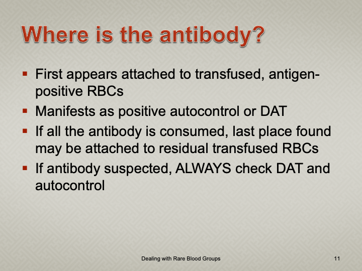
Handling Nonspecific Antibody Panel Reactions

Note: this is an updated version of a previous post.

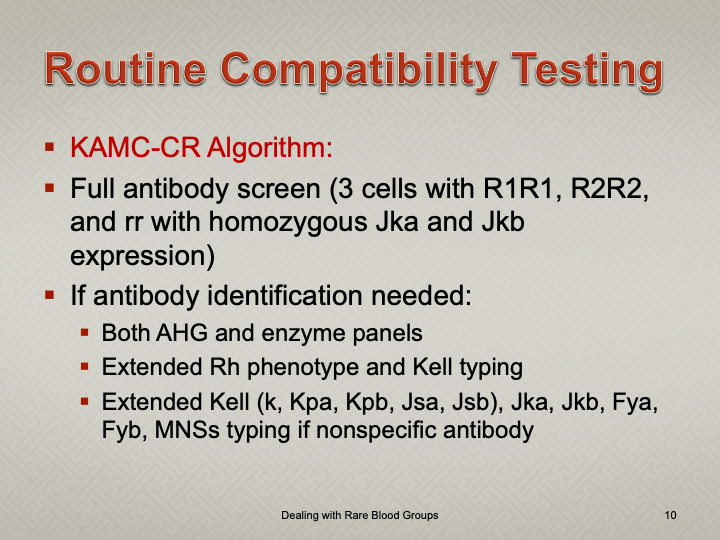
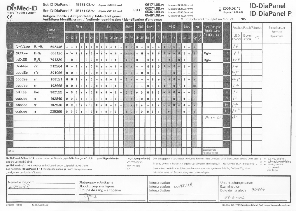
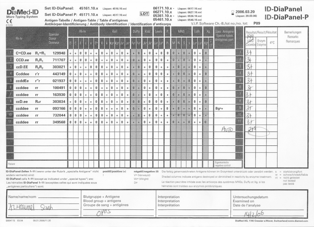
Anyone reviewing antibody panels, especially in the Middle East/Gulf region, encounters many panels for which no antibody specificity is identified.  As a transfusion medicine physician who often got called during the night for release of RBCs for patients with “nonspecific” pattern, this was a big headache.

Is it “nonspecific” because there isn’t a clinically significant antibody OR the technologist did not perform the testing or its interpretation correctly?  Does it need further testing?  Do I release blood components at this time?
In general, I do not routinely use polyspecific AHG for routine testing.  My first choice is for a gamma heavy-chain specific AHG but this is not available for gels or glass beads.  Then, I select the IgG AHG even though it does react with light chains and can detect IgM cold antibody reactions.

In general, with nonspecific reactions, I recommend the following:

  1. Repeat with a gamma heavy-chain specific reagent if the initial workup was made with another type of AHG (polyspecific or whole molecule IgG).
  2. Always do enzyme panels, sometimes with both papain and ficin reagents:  many Rh antibodies are optimally detected only at enzyme (example:  R1R1 patient with only anti-E at AHG phase but anti-E and anti-c at enzyme).
  3. Perform an extended Rh/Kell/Duffy/Kidd/Kell/MNSs/P1 phenotype and specifically check for those negative typing results AND for dosage (could the antibody only be detected in homozygous cells like many anti-M are?).
  4. Perform classical room temperature, 37C, and finally AHG phase testing.  Routinely I do not do this since antibodies not detected at 37C are unlikely to be clinically significant.  Sometimes, the AHG phase reactivity is a cold antibody of high-thermal amplitude.
  5. If the Jka or Jkb antigen typings are negative, repeat using a polyspecific AHG reagent.
  6. Use additional panels from multiple manufacturers.  Some reagents detect more nonspecific reactions than others.
  7. Try other potentiators than LISS such as PEG.
  8. Check the outdate of the panel and reagents:  if less than 1 week remaining, consider repeating with fresh reagents and getting a new patient sample.

Finally, if you still cannot define the specificity, consider repeating the testing after several days.  Maybe it is a newly emerging or an anamnestic response.

I emphasize as a physician, I do not care to see all possible antibodies present in the specimen but rather only those likely to be clinically significant.  In general, there is a shortage of labor in the blood banks so I want to eliminate unnecessary work.

Autologous Transfusion

This a revised version of a previous post for the processes of autologous transfusion that I developed at HMC Doha.  It can serve as a template for other sites and was also a teaching document for the Transfusion Committee members.

Background:

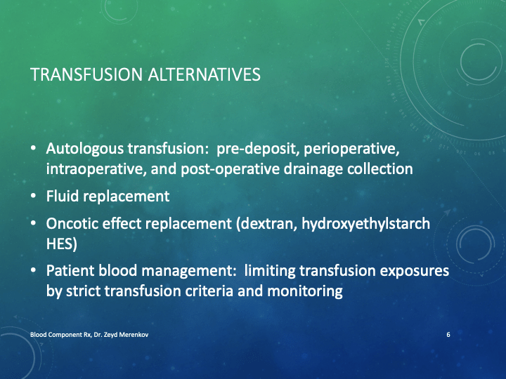
There are four basic types of autologous transfusion:  preoperative, perioperative hemodilution, intraoperative, and postoperative drainage/collection.  The use of all of the above techniques can significantly decrease the need for homologous blood and as an added benefit reduce the risk of the disease transmission and immunosuppressive effects of such homologous transfusions.

Preoperative collection can make available packed red blood cells, whole blood, platelets, FFP, and/or cryoprecipitate.  However, at most two units of blood per week can be collected.  RBC’s can be stored for up to 42 days in the liquid state, frozen RBC’s up to ten years, platelets up to five days, and fresh frozen plasma and cryoprecipitate up to one year.  The last collection cannot be less than 72 hours prior to the surgery time.  Units can be collected as long as the patient’s hematocrit remains above 33%.  Supplemental iron and erythropoietin can increase the number of units harvested.  The biggest obstacle to using this service is the coordination of the patient scheduling for this procedure.  The blood bank does not have the resources to prospectively analyze the surgical scheduling and make the various appointments, contact the attending physician, etc.  Thus, this service is vastly underutilized.

PHD or Perioperative hemodilution (also called acute normovolemic hemodilution) is useful in cases when the anticipated blood loss is at least one liter and the initial hematocrit is at least 34%.  This includes essentially all types of surgery, but in particular cardiac, vascular, orthopedic, and urologic cases.  The patient’s hematocrit Hct. is lowered to the range of 20-25% and the blood is replaced by crystalloid in a ratio of 3:1–i.e. three times as much fluid as blood, or in the case of colloid replacement, a 1:1 ratio of colloid plus 0.5 to 1.0 ml. of crystalloid.  Crystalloid has the advantage of being readily removed by diuretic use.  However, this technique should not be undertaken when vascular access is inadequate or appropriate monitoring devices are lacking.  The physician performing PHD must be familiar with the compensatory mechanisms normally invoked when the hemoglobin is acutely lowered.

Another new twist to PHD is the perioperative collection of platelets by a special attachment to a cell-saving machine.  This could allow collection of a typical apheresis load, about 6 to 10 units of fresh platelets for potential use.  There are currently studies underway to determine if this has particular clinical advantages to warrant the additional cost.

Intraoperative salvage may be performed with a number of canister or automated devices.  The latter is usually used when there are large volumes (usually 3 or more units) of blood to be salvaged.  Depending on the body site, the recovered material is at least filtered and may or may not be washed.  Care must be taken to collect the blood at a low suction rate and with minimal turbulence to minimize hemolysis.

Postoperative drainage collection of certain sites such as post-knee replacement surgery or chest wounds involves a canister collection device.  This blood may or may not be filtered before reinfusion.

Note that perioperative and intraoperative material can only be transfused up to six or eight hours at room temperature or 24 hours if refrigerated at 1-6 degrees (depending on the method used) post collection to minimize the risk of infection.  Intraoperative collection may be contraindicated in cases of cancer and if the bowel has been violated.

Other Issues:

The transfusion criteria for autologous blood is the same as for allogeneic units.

The same compatibility testing algorithm applies both the autologous and allogeneic units.

Policy:

  1. Scope:
    1. Predeposit collection of Whole Blood/RBCs and plasma is under the authority of Transfusion Medicine.
    2. Perioperative hemodilution, intraoperative cell salvage, and postoperative drainage collection is under the authority of the National Transfusion Committee in conjunction with the Departments of Surgery and Anesthesia.
      1. The Division Head, Transfusion Medicine will liaise with the clinical departments as needed.
      2. Transfusion Medicine may provide blood collection bags for perioperative hemodilution upon request.
    3. Transfusion Medicine does not receive autologous collections—perioperative, intraoperative, or postoperative drainage collection.
  2. Processes directly under Transfusion Medicine authority:
    1. Autologous collection of whole blood/RBCs (pre-deposit) for elective surgeries may be considered especially if:
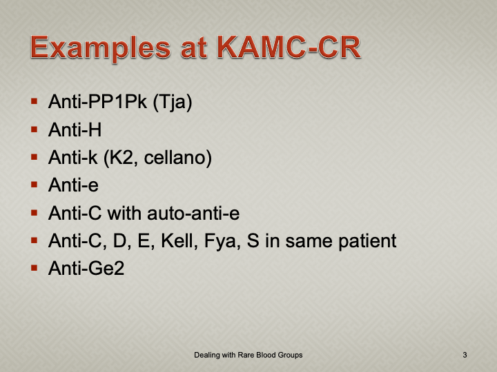
      1. The patient has a dangerous antibody for which antigen-matched units cannot be easily obtained (e.g. anti-k (cellano), anti-PP1Pk (anti-Tja), anti-H (Bombay and Para-Bombay phenotypes).
    2. Autologous collection of plasma may be considered for patients with IgA deficiency with documented specific anti-IgA antibodies.
    3. Other requests will be reviewed by the Head, Transfusion Medicine or designate.
    4. The final decision to proceed with items 2.1 and 2.2 will be made by the Head, Transfusion Medicine or his designate.
  3. Process for Transfusion Medicine Autologous Procedures
    1. The requesting physician shall provide a written or electronic order to the Blood Donor Center.
    2. The request will be reviewed by a transfusion medicine physician.
    3. If rejected, the requesting physician will be notified with the reason for the rejection.
    4. If approved, the donor shall be screened by the usual donation process except:
      1. Hgb >= 11 g/dl will be acceptable for whole blood collection.
      2. Females may also donate autologous plasma.
      3. The last autologous donation will be at least 72 hours before the elective procedure.
    5. Marker testing:  Components from autologous donors with confirmed positive cases of HBV, HCV, or HIV will NOT be used for autologous donation and will be destroyed.
    6. Computer:  Autologous collections will be entered as specifically at the time of registration in the Medinfo  Hematos IIG blood bank computer system and be labeled as autologous in their corresponding ISBT labels.
    7. The transfusion criteria for autologous units shall be the same as for homologous blood.
    8. Both autologous and allogeneic units will follow the same compatibility testing algorithm.
  4. Responsibilities:
    1. Predeposit:  Directly under the control of Transfusion Medicine for all aspects:  policies, procedures, and direct performance of the procedures, including annual review of criteria
    2. Perioperative:  Division Head, Transfusion Medicine involved in conjunction with Surgery and Anesthesia through the National Transfusion Committee.
    3. Intraoperative:  Division Head, Transfusion Medicine involved in conjunction with Surgery and Anesthesia through the National Transfusion Committee.
    4. Postoperative:  Division Head, Transfusion Medicine involved in conjunction with Surgery and Anesthesia through the National Transfusion Committee.

References:

  1. Standards for Blood Banks and Transfusion Services, Current Edition, AABB, Bethesda, MD, USA, 2014
  2. TRM.41600 CAP Checklist Standard

Opinion: Outsider Access to Blood Bank Software

I designed my blood bank software Medinfo for use by my staff at all levels and positions to adhere to and facilitate compliance to the workflow processes.  Blood bank staff were restricted to access only those functions needed for their job duties.

Blood Donation records could not be viewed by outside staff for confidentiality reasons.  Blood donor records were not linked to patient records at all.

We did not allow access for Medinfo to non-Transfusion Medicine staff since the screens were designed to maximize efficiency of the work processes, not the viewing by outsiders of results.  Outsider access was made through the hospital information system HIS.

The separate hospital information system HIS interfaced to Medinfo for the following functions:

  • Ordering blood components

  • Ordering a limited number of tests from which algorithms would be generated on the blood bank side for further testing
  • Querying the status of the test or component orders (e.g. ordered, collected, in blood bank being processed, completed)
  • Viewing of completed tests and component requests

Even within the HIS ordering capabilities, there were additional restrictions:

  • Blood components:  outside doctors could order base blood components could be ordered, but special processing such as washing or irradiation followed internal blood bank rules.  Outside physicians could state their preferences in an order comment, but blood bank rules applied.  Any disagreements had to be discussed with the transfusion medicine physician.
  • Testing:  Only base tests such as ABO/D typing, antibody screen, direct antiglobulin test, transfusion reaction workup, and cord blood testing could be directly ordered by outside physicians, but further testing depended on the results of these tests as allowed by internal blood bank algorithms.  An outside physician could not directly order other tests but had to discuss his concerns with the transfusion medicine physician.

Results viewing in the HIS were subject to additional conditions as well:

Only certain results, not all results were viewable directed in the patient’s chart since showing all results may be confusing to the outside physicians and nurses.  The selected results were sent back into the HIS for viewing.  All these non-viewable results were retrievable for blood bank staff in Medinfo.

Another option, one I did not use at either HMC Doha or NGHA in Saudi Arabia, was to order tests and components by physicians directly into Medinfo.  Likewise, they could view test results directly in the system.  Special screens could be constructed to offer ordering and results retrieval.

Use of Expired Reagents
This is a sample document for use of expired reagents I wrote for HMC Qatar.

Principle:

Due to logistics issues including the long distance between suppliers in Europe and North America and Qatar and the importation/customs clearance of critical materials, Transfusion Medicine has developed a contingency variance policy to minimize disruption of the essential transfusion medicine testing and component preparation.  Approval for use of outdated reagents in special circumstances is not meant to be an excuse for untimely monitoring and improper ordering of supplies.

Definition:

Rare Reagent:  Any reagent that is either used uncommonly or is in short supply and difficult to obtain in a timely matter.

Policy:

  1. Maintain a minimum six (6) months’ supply of each reagent when feasible.
    1. This cannot be done for short-outdate reagents such as reagent red cells for panels and antibody screens.
  2. If an in-date reagent is not available for use, then an outdated reagent may be considered for use if:
    1. It passes an in-run quality control including negative and positive controls as applicable specific for the test in question.
    1. Supervisor reviews the results and approves their use:
      1. Results using such outdated reagents may only be used if they pass the validation rules in that procedure.
    1. The Division Head, Transfusion Medicine, or designate reviews the supervisor’s recommendations and approves their use.
  3. All such variances must be documented as follows:
    1. Variance document form
    1. In the comments for results within the Medinfo Hematos IIG software.

References:

Sections 1.3.2 and 7.0, Standards for Blood Banks and Transfusion Services, Current Edition, AABB, Bethesda, MD, USA

COVID-19 Convalescent Plasma CCP Product Issue

This is the conclusion of a continuing series of posts on the actual Medinfo design of the CCP donation and release processes and covers the transfer of completed units to the hospital blood banks.  It highlights specific changes made for the parallel CCP system I developed at HMC Doha.

A blood component is either located at a production site, a destination hospital blood bank site, or in transit.  Here a quarantine production site is specified.  The actual release process is documented in this post.

In summary, with the exception of the donor marker testing and immunohematology testing, all other CCP processes are handled by special quarantine processes.  There are abbreviated marker testing specific for plasma and a special Predonation screening to minimize wastage of the expensive apheresis kits.

Weak D Phenotype

I prepared this policy for HMC Qatar, which did not perform genotyping.

Principle:

Weakened expression of the D antigen may be due to either quantitative or qualitative defects.  Qualitative defects (partial or mosaic D) are uncommon.

Patients with quantitative defects have the whole D molecule and will not make anti-D upon exposure to D-positive RBCs.

Patients with partial or mosaic D may form antibodies against the portion of the D molecule they lack.  However, not all partial D types may make anti-D upon exposure.  Since we cannot specifically test for this condition, we shall consider females of child-bearing age with weak D as potentially partial D and use D-negative RBCs.

Genotyping for the D gene may define better which partial D types need to receive RhIG or D-negative transfusions.

Definition:

Weak D is defined as weak (1+ or weaker) reactions with one or more routine reagents or reactions only at antiglobulin phase (i.e. Du testing).

Policy:

  1. All donor units must be tested by a method to detect weak D phenotype.  If such testing is positive, the donor unit must be labelled as D-positive and be used routinely for D-positive patients.
  2. Weak-D females of child-bearing ages (<50) should receive D-negative RBCs.
  3. Weak-D males may receive D-positive RBCs unless they are a potential recipient of chronic transfusions (e.g. thalassemia, sickle cell disease, etc.)

References:

Technical Manual, Current Edition, American Association of Blood Banks, Bethesda, Maryland, USA

Standards for Blood Banks and Transfusion Services, Current Edition, AABB, Bethesda, MD, USA

Issues with Labels

You can have the most sophisticated blood bank software, but if you can’t read the labels or if they fall off, you have a disaster.  These are my thoughts from implementing our blood bank computer system back in 2013.

Check Digits:

Both ISBT specimen and product labels have an internal system to verify that they have been read correctly.  Within the blood bank software, this should not be a problem.  However, can your third party such as a hospital information system HIS read them?

ISBT Compatibility:

The institution’s HIS could not read the component labels.  To this date, the problem has not been fixed.  As a workaround, we sent them the ISBT label codes directly from the blood bank software.  The only complete transfusion record was in the dedicated blood bank computer system, not the HIS.  You could not rely on the bedside nursing entry at all.

The HIS did not use the ISBT database and had no values for the E codes.  Again, we had no choice but to send an abbreviated E-code descriptor to them.  We did not use their transfusion module at all, but one of our clients did.  They had to hard code the list of E codes in use with their descriptors into their HIS.

Label Adhesive

We tested candidate labels at room temperature, 1-6, minus 18, and minus 80 C.  We found that most of the labels’ adhesive were not sticking at minus 80.  For some, you could literally blow on the blood bag and the label fell off.  I imagined a scenario in which I opened a freezer and saw the blood bag labels all lying separately at the bottom.

RFID Tags:

Do you use an RFID tag integrally attached to the ISBT label OR do you stick a separate RFID tag?  If the latter, how do you ensure that you put the proper tag on the proper bag?

Readability:

Readability:  Can all your blood bank devices read your printed labels?  Do you have to adjust the printers for this?  Whose responsibility is it to do this? 

Labels printed outside the blood bank:

If you receive patient specimens from outside the blood bank, can your devices read them?  Who is responsible to adjust the printers in the wards and clinics?

Validations:

Who validated that the HIS prints the accurate complete label for the right patient?  We discovered that this was not the case with our HIS and needed correction by them.  Remember that any processes affecting Transfusion Medicine should be assessed by Transfusion Medicine.  Do not accept verbal assurances from anyone, not even your HIS.

2/2/21

COVID-19 Convalescent Plasma CCP Interdepot Transfer

This is a part of a continuing series of posts on the actual Medinfo design of the CCP donation and release processes and covers the transfer of completed units to the hospital blood banks.  It highlights specific changes made for the parallel CCP system I developed at HMC Doha.

A blood component is either located at a production site, a destination hospital blood bank site, or in transit.  Here a quarantine production site is specified.  The actual transfer protocols and allowable destination sites are listed for this product.