Post RhIG Use Without Antibody Identification

Rh immunoprophylaxis has big effect on the Transfusion Medicine operations.  With a very active obstetrics program, there are numerous prenatal blood bank workups and administrations of Rh immune globulin RhIG—and consequently many antibody identifications.  In fact at my previous position, the largest single source of antibody workups was from patients post-RhIG administration.

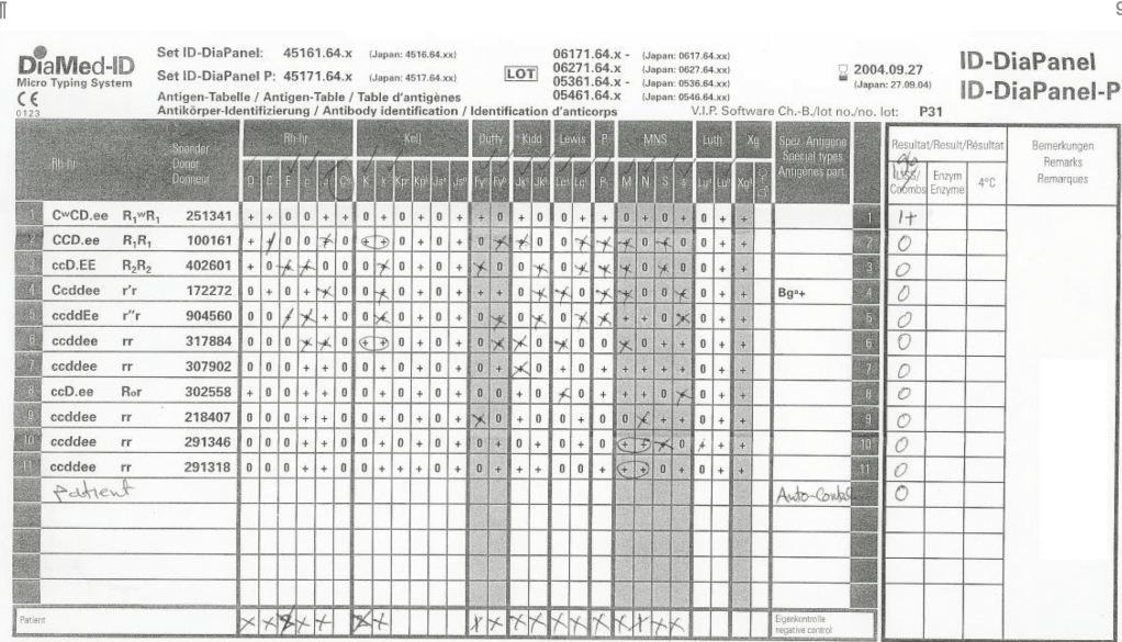
As a result of the antenatal RhIG use, there were numerous positive antibody screens.  Our policy had been to always do a full antibody identification, both AHG and enzyme panels, and this made up the bulk of our antibody testing.  In my experience, I have found other antibody specificities in some of these patients, including anti-Kell and anti-c, both of which could be very clinically significant.

As part of the workup, we also did ABO and extended Rh and Kell phenotyping, and thus identified rare Rh phenotypes (r’r’, -D-, etc.) and several Bombay phenotypes.

Unfortunately, I have been at institutions where they assumed a positive antibody screen in a patient with recent RhIG administration was passive anti-D and did NOT perform further testing.

If the patient needs blood and the antibody screen is positive, the antibody workup must be performed for routine release.  This means about a 20-30 minutes delay in release until the antibody panels are completed and reviewed.  Otherwise, blood can be released through an emergency protocol if the clinician accepts responsibility for incomplete testing at the time of release.

Many clinicians refused to do this emergency release and insisted on waiting for the antibody identification before accepting RBC components.  They would not take the responsibility for the emergency release.

If we had done the antibody workup in these RhIG patients, we would have identified the anti-D, and I as the transfusion medicine physician would then advise the clinician to accept emergency release of RBCs while we complete the antibody workup.

For this reason alone, I insisted on a full antibody workup for all RhIG patients RhIG who exhibit a positive antibody screen.  I would be comfortable in recommending emergency release if I had results of the previous antibody identification showing passive anti-D.  The delay in release could adversely affect the patient’s outcome if there is active bleeding.

In summary, if a patient has a positive antibody screen post RhIG administration, you should still do ABID to rule out other antibodies and facilitate release of RBCs to a bleeding patient.

Process: Donor Collection

Process:  Donor Collection

Zeyd Merenkov, MD, FCAP, FASCP

Independent Consultant in Transfusion Medicine

5.4.1 PROCESS DONOR COLLECTION:

Process:

  1. Donors must pass and complete all previous processes in the donor workflow (registration, questionnaire, and physical examination) before the collection process begins.
  2. The donor is positively identified by a designated picture ID and Hematos donor consent form with specimen/encounter number and barcode.
  3. Donor staff checks and prepares a suitable vein
  4. Donor staff collects/labels specimens and the whole blood or apheresis components AT THE DONOR’S BEDSIDE.
  5. Donor reactions are assessed and treated as they occur.
  6. Donors are observed in a post-donation area and given post-donation instructions before discharge.
  7. All processes are documented in Hematos IIG.
  8. Donor units and specimens are sent to component processing and donor marker testing.
  9. The collection workstation and equipment are cleaned before starting a new donor collection.

References:

  1. HMC 1001 Setting Specification, Version 1.5, Hematos IIG, Medinfo
  2. Standards for Blood Banks and Transfusion Services, Current Edition, AABB, Bethesda, Maryland, USA

Policy: Donor Collection

5.4 POLICY: DONOR COLLECTION

Policy:

  1. All donors will be positively identified with a picture ID and by their Hematos identifiers (donor ID and session registration/specimen number).
  2. All previous processes (registration, donor deferral database check, questionnaire, and physical examination) must pass.
  3. The donor’s arm veins will be inspected for a suitable donation site and prepared by a suitable aseptic technique.
  4. Whole Blood:
    1. Only whole blood units collected within the specified time interval may be used for component processing.
  5. Apheresis:
    1. Apheresis units will be collected at frequencies to keep the total RBC loss below 200 in any 8-week period.
    2. Only apheresis units collected within the specified time interval may be used for component processing.
  6. Donors will be treated for adverse reactions as needed.
  7. All specimens and donor units will be labeled at the donor’s bedside before starting a new donor collection.
  8. Donors will be monitored post-donation for a reasonable interval before discharge.
  9. All processes will be documented in the Hematos blood bank computer system.
  10. All equipment and supplies will be used according to manufacturer’s instructions.
  11. The collection workstation and equipment will be cleaned before starting the next donor.
  12. All policies, processes, and procedures must comply with Qatari, HMC, and applicable accreditation standards (i.e. AABB, CAP, and JCI).

References:

  1. HMC 1001 Setting Specification, Version 1.5, Hematos IIG, Medinfo
  2. Standards for Blood Banks and Transfusion Services, Current Edition, AABB, Bethesda, Maryland, USA, October 2013

How I Select Technologist Candidates for Work or Promotion

As a transfusion medicine physician, I must know if I can trust my staff’s interpretation of immunohematology testing.  I may be called at night and they will provide me with results and I must use these to make a medical judgment.  If their interpretation is flawed, I might make a decision that harms the patient.

I really don’t like multiple-choice questions, but nowadays this is often the norm.  For my staff, especially senior staff and those who want to be promoted to senior staff, I have developed a series of projective exercises to help me understand their thought processes. I also used this approach on prospective candidates for hire.

Here is a sample exercise.  I have the candidate or staff review this panel and tell me to interpret it:

Most of them answer that this is an anti-Cw without hesitation.  However, they are basing that on only one Cw-positive cell.

More astute ones indicate it might be anti-Cw but ask to test additional Cw-positive cells and perform an enzyme panel.  These are the ones that I will consider for hire or promotion now.

Product Delivery in Medinfo 1

This is the start of a series of posts on how Medinfo blood bank software was designed for product delivery in the HMC system in Doha.

The overall process was:

  1. Transfer blood components (all types) from the Blood Donor using the Interdepot Transfer process (see that post for details) to the Hamad General Hospital HGH General Delivery Deposit.
  2. Release components to individual HMC system hospitals and client blood banks from the HGH General Delivery Deposit,.

It was also possible to release blood components directly from the Blood Donor Center to HMC hospital blood banks as a contingency.  Client hospitals outside the HMC system still had to obtain their components from HGH General Delivery Deposit.

Interfaces with Quantitative and Qualitative Results

Processes and Software Building Part 7

This is an update of a previous post.

Blood Bank instruments may perform tests and release test results in a numerical or alphanumeric format or both.  For example, nucleic acid and enzyme immunoassay may release a qualitative result (e.g. positive, reactive, borderline/gray-zone, negative, nonreactive).  Alternatively, the machine may release the signal to cutoff ratio (S/CO) as a numeric result.

Blood bank software may use either kind of result on which to base interpretative rules for acceptability of the donor.  The qualitative result criteria are based on the quantitative SC/O but the equipment automatically interprets this.  The S/CO ratio of 1 is the cut-off point.  Thus a value of 0.99 is negative and the value 1.01 is positive.  But is it really so clear-cut since the difference between the two is so small?  Thus, some people have added the term gray-zone for values close to but below the cutoff.  Could a value of 0.95 be an early infection?

I personally prefer to see the actual cutoff but use the manufacturer’s criteria for interpretation.  As a physician, it is good to review the S/CO on serial exams.  If a borderline or gray-zone result becomes positive, then perhaps the original result indicated early infection.  The question still remains, what is the gray-zone?  0.95 to 0.99, 0.90 to 0.99, etc.  Some accrediting schema have not used gray-zone for interpretation.

With Medinfo’s blood bank software, I could choose either option or both—or at least store the S/CO as a nonreported result for subsequent review.  I could even chose, test by test, in a series between reporting either S/CO or the qualitative result.

Semiquantitative results, e.g. in {0, 1+, 2+, 3+, 4+} are qualitative and could also include mixed field (mf) and hemolyzed (h).  I showed examples of this with ABO/D antigen typing in a previous post—see attachment.

On the contrary, the results from blood production equipment may include parameters such as time of preparation, original volume, final volumes for each component, platelet yield index as an indirect measure of platelet count.  When there is pooling, the final total volume is critical to determine if pathogen-inactivation procedures and platelet additive solution can be used.  This is a much more complicated interface.

Process: Donor Physical Examination

This is a sample process document for donor physical examination. Compare it to the previous post Policy: Donor Physical Examination.

5.3.1 PROCESS DONOR PHYSICAL EXAMINATION:

Process:

  1. The donor is positively identified by a designated picture ID and Hematos donor consent form with specimen/encounter number and barcode.
  2. Donor staff measures vital signs of donor and enters results into Hematos IIG.
  3. Donor staff inspects donor arms for suitable veins and checks for concurrent skin diseases and/or scarring.
  4. Medinfo Hematos IIG determines donor eligibility to collect components.

References:

  1. HMC 1001 Setting Specification, Version 1.5, Hematos IIG, Medinfo
  2. Standards for Blood Banks and Transfusion Services, Current Edition, AABB, Bethesda, Maryland, USA

Policy: Donor Physical Examination

This is another example of a short concise policy statement. The process example will follow:

5.3 POLICY: DONOR PHYSICAL EXAM

Policy:

  1. All donors will be positively identified with a picture ID and by their Hematos identifiers (donor ID and session registration/specimen number).
  2. The donor’s vital signs (BP, pulse, temperature, respiratory rate) will be measured by designated Donor Center staff.
  3. The donor’s arm veins will be inspected for a suitable donation site and evidence of scarring.
  4. Donor eligibility for actual collection will be determined by the Hematos IIG algorithms based on this data.
  5. All policies, processes, and procedures must comply with Qatari, HMC, and applicable accreditation standards (i.e. AABB, CAP, and JCI).

References:

  1. HMC 1001 Setting Specification, Version 1.5, Hematos IIG, Medinfo
  2. Standards for Blood Banks and Transfusion Services, Current Edition, AABB, Bethesda, Maryland, USA

Antiglobulin Reagents

Antiglobulin reagents are used to detect molecules bound to the RBC surface.  What they detect depends on their specificity.  Such detection can be performed in the routine immunohematology laboratory or elsewhere such as flow cytometry.  This discussion is for the blood bank laboratories, both routine and reference immunohematology.

This is how I classify and use the various reagents in my daily practice:

  • Routine use—DAT testing and compatibility testing
  • Antibody identification
  • Drug-related hemolysis and transfusion reaction workups
  • Special assays:  DAT-negative AIHA, prediction of clinical significance of reaction by IgG subclass determination

There are many types of antiglobulin reagents:

  • Polyspecific:  IgG and complement usually C3d but sometimes C3b specificity included
  • Whole Molecule IgG including mu heavy chains and kappa and lambda light chains
  • Monospecific gamma heavy chain, mu heavy chain, alpha heavy chain, C3c, C3d, C3b
  • IgG subclass:  IgG1 IgG3

Whole molecule IgG detects class-specific mu heavy chains AND light chains kappa and lambda.  Since kappa and lambda are found on all immunoglobulin classes, whole molecule reagents can detect IgM so there may be weak staining with cold antibodies that are not clinically significant.

C3d is the final breakdown product of C3b and does not cause hemolysis.  Its presence merely means that at some time—unspecified—complement was fixed.  C3c is an intermediate product in the breakdown pathway.  If detected, C3c positivity means ACTIVE complement fixation was occurring at the time of specimen collection.

General Use:

Routine DAT testing:

  • Polyspecific:  if positive, then use
  • Monospecific IgG and monospecific C3 reagents

Antibody workups:

Routine:  antibody screens and AHG crossmatch (if indicated)

  • Polyspecific
  • Gamma heavy-chain monospecific

Monospecific gamma heavy chain is preferred to minimize non-clinically significant, cold antibody interference.

Complicated—where detection of complement reactivity is especially important:

Polyspecific for drug-related hemolysis and transfusion reactions

Difficult antibody workups, e.g. to rule out anti-Jka and/or anti-Jkb

Specialty Reference Procedures:

  • DAT-negative AIHA:  mu heavy chain, alpha heavy chain for IgM and IgA mediated hemolysis (rare), C3c to detect active complement fixation
  • Predicting clinical significance:  IgG1 IgG3

Complement fixation may be important in various drug-related hemolysis and some transfusion reactions so I always use a polyspecific reagent in these situations.  Most antibodies can be detected by gamma heavy-chain specific reagents;  however, there are rare examples of anti-Jka and anti-Jkb which are only detected by complement.  Whenever I have a nonspecific reaction in an Jka-negative or Jkb-negative patient, I repeat the AHG panel using polyspecific reagents.  I do not use polyspecific routinely because of the nonspecific and non-clinically significant cold antibodies.

One way to assess for the clinical significance of an antibody is to determine its IgG subclass.  In general, IgG3 antibodies may fix complement and cause severe hemolysis.  Both IgG1 and IgG3 antibodies cross the placenta and may cause hemolytic disease of the fetus/newborn.

In summary, when reviewing immunohematologic reactions using AHG, I always remember to check which type of AHG reagent was used.  I always keep multiple types of AHG reagents in the laboratory for the reasons explained above.