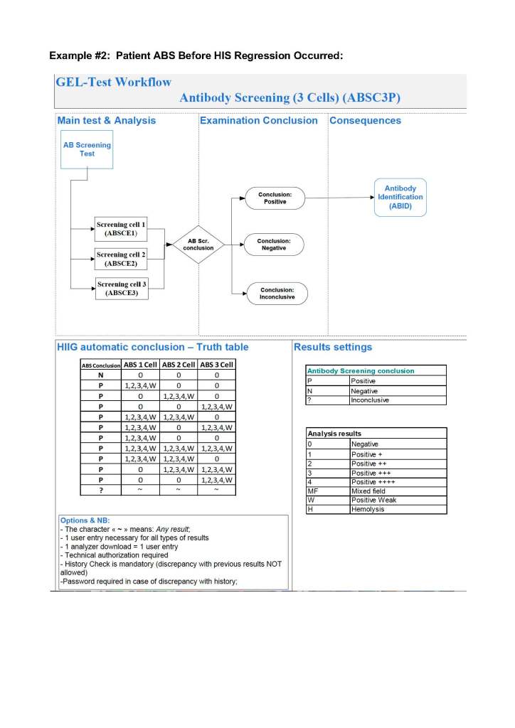
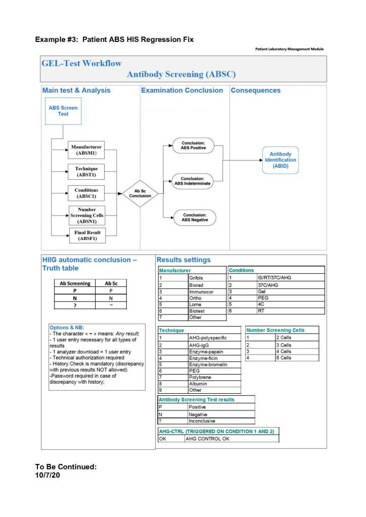
Data Entry Verification: Updated Version

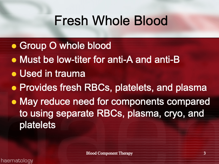
This is an update from the previous version posted to include low-titer group A FFP/FP24 and low ABO-titer group O whole blood.

Principle:

This policy outlines steps taken to minimize the risk of data entry errors and is based on a dualistic approach:  review of results by a senior technologist and/or supervisor and various computer safeguards built into the Medinfo Hematos IIG blood bank computer HIIG system.  This policy also discusses the verification (here called authorization) and purge processes of HIIG.

Policy:

  1. Review by senior technical, supervisory, or transfusion medical staff:
    1. Designated test procedures require review by a second technologist before authorization.
    2. Complex immunohematology testing and specimens showing aberrant results (e.g. ABO/D discrepancies) are reviewed by the supervisors or designates and ultimately a transfusion medicine physician before authorization.
  2. Computer system HIIG rules:
    1. Privileges:
      1. System restricts which staff can perform specific tests
    2. Patient/donor identity:
      1. System asks end-users to verify patient/donor identity before starting any access to the patient/donor record.
      2. System performs historical database checking and flags any inconsistencies (e.g. historical ABO/D typing differences, etc.)
    3. Testing:
      1. Only selected staff have privileges to authorize or purge.
      2. ABO/D testing algorithms require entry of reactions, not interpretation of results and are compared to a truth table.
        1. Aberrant results require special review before ABO/D typing results can be authorized/purged.
        2. D-controls must be negative to allow D typing results to be authorized for liquid D-typing reagents.
      3. DAT results require appropriate controls to meet truth-table criteria.
      4. Eluates require last wash to be negative before authorization
    4. Blood components:
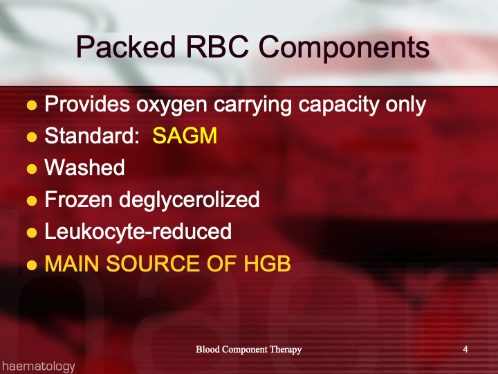
      1. Selection of RBC or plasma units requires two independent sample determinations within 72 hours of each other.
      2. ABO-incompatible RBC or FFP/FP24 transfusions are not allowed.
      3. Titer-based ABO blood group selection:
        1. Low titer group A FFP may be used as universal plasma like group AB.
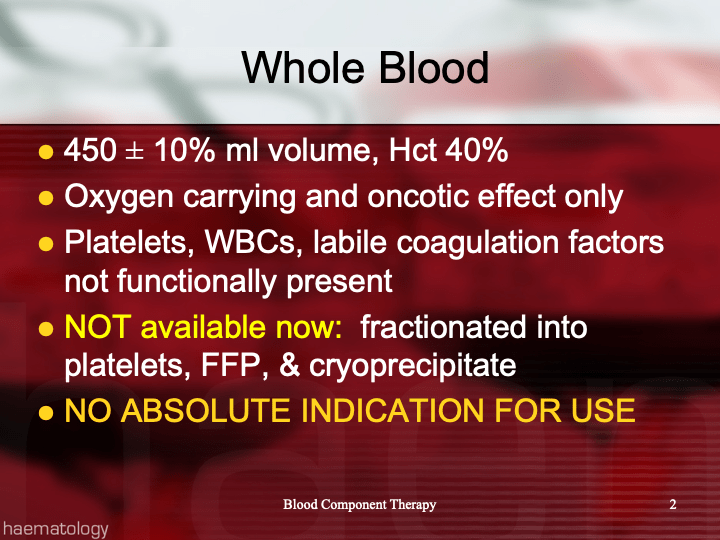
        2. Group O whole blood with low anti-A and anti-B titers may be used for all ABO types.
        3. Acceptable titer threshold is specifically defined as parameters in Medinfo.
      4. Donors with any detectable clinically significant antibodies are permanently deferred.
      5. Depending on the patient’s antibody history, release of RBC units may require antigen-matched units.  Examples:
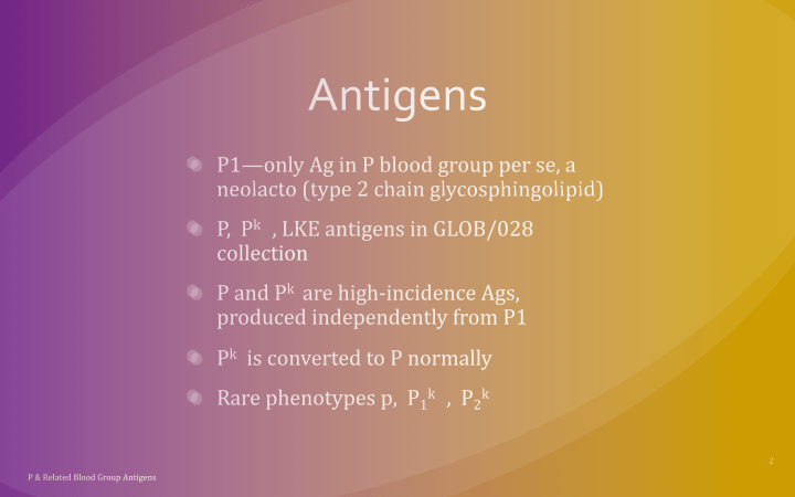
        1. Mandatory matching (only antigen negative matched units allowed—no antigen positive or antigen-untyped units):  Antibodies against H, D, c, K, k, Kpa, Kpb, Jsa, Jsb, Jka, Jkb antigens, anti-PP1Pk
        2. Priority matching (incompatible or untested can be approved by a transfusion medicine physician):  C,E, e, Fya, Fyb, M, S, s
        3. Antigen matching not required:  Lea, Leb, N
      6. Least-incompatible crossmatch require special authorization to release
      7. Protocols to force irradiation or other modified components can be setup in HIIG.
    5. Donors:
      1. Donor tests have same criteria as the same test used in patient testing for controls, etc.
      2. Donor demographics are read directly from the Ministry of Interior database—no manual entry (bar code only used).

References:

  1. Workflows for Hematos IIG (1001 through 1005), 2013-2020
  2. Standards for Blood Banks and Transfusion Services, Current Edition, AABB, Bethesda, MD, USA
  3. Guidelines to the Preparation, Use, and Quality Assurance of Blood Components, European Committee (Partial Agreement) on Blood Transfusion (CD-P-TS), Current Edition

Preventing Graft vs. Host Hemolytic Anemia

Principle:

Donor lymphocytes in an organ transplant may make antibodies and cause a clinically significant hemolytic anemia, i.e. a graft vs. host hemolytic anemia GVHHA.  Optimal handling in these cases should include antibody screening/identification for all potential donors and recipients.  The transfusion medicine physician should review the results for possible issues of antibody/antigen incompatibilities to proactively select matched blood components and avoid GVHHA.

In the Medinfo blood bank computer system, we can make custom rules to ensure release of only antigen-matched units as needed.

Policy:

  1. Perform antibody screen and identification (if indicated) for all prospective organ donors and recipients.
  2. If the organ donor has clinically significant antibodies, check if the recipient has the corresponding antigens.  If so, select RBC units negative for the donor antibody specificities. 

Example:  Donor has anti-Kell (K1) and patient is K1-positive.  Use only K1-negative RBCs post-transplant.

  • Send the case to the transfusion medicine physician to review.  He will contact the clinicians as indicated.
  • Create a rule in Medinfo forcing the antigen matching.

References:

  1. Technical Manual, Current Edition, Bethesda, MD, USA
  2. Standards for Blood Banks and Transfusion Services Current Edition, AABB, Bethesda, MD, USA

Patient D-Typing Algorithm Using Ortho Monoclonal Cocktail Reagents

Principle:

We must select D-negative RBC units for transfusion if the patient is truly D-negative or if he/she is a partial D since transfusion of a partial D positive unit may induce antibodies against any part of the D molecule.  Thus, for patients, we will consider patients with partial D as D-negative.  Note that this is NOT the usual practice in the USA;  however, AABB Standards do allow that we do NOT test patients for weak D and give D-negative RBCs instead.

Background:

Ortho Diagnostics Reagents use two different monoclonal antibody cocktails that react variably with the antigen D (Rh1)—these are found on the card:  Anti-A/B/A,B/D/D/Ctrl:

Anti-D/Anti-RH1—IgM monoclonal antibody clone D7B8 can detect most examples of weak and partial D including weak D types 1, 2, 3, 4.0, and D categories II, III, IV, V, VII, DBT, and R0HarIt does NOT detect category VI.  Retest positive reactions of 2+ or less by an alternate method.

Anti-D/Anti-RH1—IgM monoclonal antibody RUM1 can detect most examples of weak and partial D including weak D types 1, 2, 3, 4.0, and D categories II, III, IV, V, VII, DBT, and R0HarIt does NOT detect category VI.  Retest positive reactions of 2+ or less by an alternate method.

Policy:

  1. Follow the manufacturer’s instruction for storage, handling, and usage of all reagents.
  2. If the D-control is positive, the reactions are indeterminate, repeat by another method.
  3. Run both anti-D reagents listed—do not use the donor typing algorithm or reagents.
  4. Use the following table for interpretation and further actions if needed:
Pattern #Anti-D/D7B8Anti-D RUM1D-Interpretation
1PositivePositiveD-positive
2PositiveNegativeDo additional testing
3NegativePositiveDo additional testing
4NegativeNegativeD-negative  

If the reaction is 2+ or less with either the Ortho anti-D/D7B8 reagent or anti-D/RUM1 or if the patterns 2 or 3 above, repeat by another manufacturer’s reagents.  In the meantime, consider the patient as D-negative.

Medinfo-Ortho interface settings for Patient Testing:

Anti-D/D7B8Anti-D RUM1D-Interpretation
3, 43, 4D-positive
3, 40~
03, 4~
00D-negative
~~~

Note all of the following:

  1. If the result is D-indeterminate, use D-negative RBCs.
  2. No reagents may be able to detect all D variants.
  3. ~ means any other value for that reagent (1+, 2+, mf, hemolyzed)
  4. Note that this new algorithm makes a 2+ reactivity as indeterminate with Anti-D/D7B8 or Anti-D/RUM1.

References:

  1. Publication e631300291, Product Insert, Anti-A/B/A,B/D/CDE/Control Card, 2010, Ortho Clinical Diagnostics, High Wycombe, Buckinghamshire/UK
  2. Standards for Blood Banks and Transfusion Services, Current Edition, AABB, Bethesda, MD, USA

Information to Collect Before Contacting the Transfusion Medicine Physician

The Transfusion Medicine Physician needs certain minimal information to make a medical/clinical assessment and decide what action needs to be taken.

Policy:

  1. Before contacting the transfusion medicine physician, please obtain the following:
    1. Clinical diagnosis
    2. Transfusion history
    3. Medication history
  2. In the case of transfusion reactions, follow Interim Policy Transfusion Reaction Workup and be certain to include the following information before contacting:
    1. Vital signs (BP, pulse, temperature, respiratory rate) both pre- and post-transfusion
    2. Clinical symptoms (e.g. rash, urticaria, fever, respiratory distress, etc.)
    3. Pre-transfusion DAT if post-transfusion DAT is positive.
    4. Donor Unit DAT and eluate on the post-transfusion specimen if the post-transfusion DAT is positive and pre-transfusion negative.
    5. Eluate on the post-transfusion specimen if both the pre- and post-transfusion DAT are positive.

6/11/20

Rh Immune Globulin Candidacy Triage

Principle:

All D-negative women without immune anti-D must be screened for the need to receive Rh immune globulin as soon as possible after birth.  If the mother is determined to be a candidate, then her specimen must either be screened for a large fetomaternal hemorrhage (FMH—e.g. by the E-rosette or similar technique) or directly quantitated for FMH by either the Kleihauer-Betke or flow cytometry.  The whole process must be completed so that the mother receives the entire dose of RhIG within 72 hours after delivery.

Pregnant women should have an ABO/D typing early in the pregnancy and an antenatal dose between 26-32 weeks.  Thus, at the time of birth, passive anti-D may be detected.  It is essential to know the history of RhIG administration.  In the following example, RhIG is dispensed by the Pharmacy.  If the RhIG is released by the blood bank, records should be easily obtained.

This procedure covers the hospital blood bank processes.  Close coordination between Transfusion Medicine and the obstetricians is essential for a successful Rh immunoprophylaxis program.

Abbreviations:

RhIG:  Rh immune globulin

FMH:  Fetomaternal hemorrhage

All plasma derivatives, including RhIG, are dispensed from Pharmacy.  It is the responsibility of the blood bank technologist to check the Pharmacy records for the history of RhIG administration.

For immunoprophylaxis after accidental Rh(D)-incompatible RBC transfusions, see the separate interim policy by that name.

Policy Details:

  1. Mothers with active (immune) anti-D are excluded from this procedure.  They do NOT need RhIG and to give them it is a waste of a valuable resource.
    1. If the D-negative patient has other RBC antibodies but no anti-D, then she remains a candidate for RhIG prophylaxis.
    2. RhIG has no effect for preventing alloimmunization to other RBC antigens.
  2. D-negative mothers with first-trimester abortions should be given at least 50 mcg IM RhIG dose.
    1. If clinically a large fetomaternal hemorrhage is suspected, then screen/quantitate for fetomaternal hemorrhage as detailed below.
  3. If the D-negative mother undergoes amniocentesis, version, or has had abdominal trauma, the obstetrician may request RhIG or if he suspects significant hemorrhage, fetomaternal hemorrhage quantitation and calculation of the RhIG dose as indicated below.
  4. Platelet transfusions from Rh(D) positive donors:
    1. Platelets derived from a Rh(D)-positive donor prepared by the buffy coat method or by apheresis, either suspended in plasma or PAS do not require RhIG administration.
    2. If a D-negative mother receives legacy, platelet-rich-plasma-derived platelets from a D-positive donor, it is at the discretion of the clinician to request one 300 mcg vial IM RhIG as potential immunoprophylaxis.  (Note:  PRP platelets are no longer prepared at HMC.)
  5. The cord blood of all infants from D-negative mothers without immune anti-D must be typed immediately for the D antigen upon receipt in the Blood Bank.
  6. If the D-negative mother received antenatal RhIG or if it is unclear if the anti-D detected in her blood is passive (i.e. antenatal) or active (immune), she must still be screened by this procedure.
  7. If the baby is D-negative, no further processing is necessary:  it is not necessary to give RhIG.
  8. If the baby is D-positive or if its D type is unknown, a maternal sample (EDTA-anticoagulated blood) must be screened for FMH (e.g. by the E-rosette technique).  This is done in Hematology Laboratory.
    1. The E-rosette test will detect a minimum of 10 ml of fetal RBCs.
  9. If the screen is not available, then direct FMH quantitation must be done (e.g. by either the Kleihauer-Betke staining or flow cytometry).  This is done in Hematology Laboratory.
  10. If the FMH screen is negative, then the mother should receive one 300 mcg. standard vial of IM-RhIG.
  11. If the FMH screen is positive, the dosage of RhIG must be determined by the amount of fetal bleed (expressed as “ml of fetal bleed”.)
  12. All FMH quantitation results must be calculated by a transfusion medicine physician or by the clinical service physician responsible for patient’s care and the dose of RhIG must be calculated as follows:
    1. Verify the type of RhIG available in Pharmacy:
      1. Type: IV or IM
      2. Dosage:  240/300 mcg and manufacturer’s suggested protection level:
        1. Each 240 mcg IM usually protects against 12 ml fetal bleed
        2. Each 300 mcg IM usually protects against 15 ml fetal bleed
        3. Note:  dosage depends on the origin of the pharmaceutical material.
    1. Calculate the number of vials as follows:
      1. Blood Volume in ml = (Weight in kg) X 70 ml/kg
      2. RBC Volume in ml = (Blood Volume in ml) X Hematocrit
      3. Fetal Bleed in ml = (%Fetal Bleed) X (RBC volume in ml)
      4. # Vials 300 mcg IM RhIG preparation = (Fetal Bleed in ml)/15
      5. # Vials RhIG 240 mcg IM = (Fetal Bleed in ml)/12
      6. Note:  some would add 1 additional vial to this calculation.
  13. If the calculation is performed by the Transfusion Medicine Physician, he should append a comment to the postpartum antibody screen ABID result of the mother using Medinfo Hematos IIG.
    1. If the calculation is performed by a designated individual outside Transfusion Medicine, he should document it in the patient’s medical record.
  14. The appropriate RhIG dose should be administered within 72 hours of delivery whenever possible.
    1. If greater than 72 hours post-delivery, still administer the dosage; however, the efficacy at preventing Rh(D) alloimmunization may be reduced.

Note:

  1. RhIG will only protect against forming anti-D antibodies, not any other clinically significant antibodies.
  2. If the patient is D-negative but has other clinically significant antibodies, still administer the RhIG as per the protocol above.

References:

  1. Technical Manual, Current Edition, Bethesda, MD, USA
  2. Standards for Blood Banks and Transfusion Services Current Edition, AABB, Bethesda, MD, USA

Opinion: Understanding Each Step of the Process

During an AABB inspection many years ago, I observed the Lead Assessor interacting with a medical technologist at the bench.  As expected, the Assessor asked in detail about the test and where the documentation and any teaching aids were located.  Most importantly, the tech was asked to explain the procedure step-by-step and if they knew WHY each step was done and how the various outcomes affected the test.

In my opinion, the WHY is the most important issue.  In our testing and processing, we are not baking a cake.  We do not need a mindless automaton to perform the steps without knowing what they are doing.  To get it right, the technologist must understand why he/she does each and everything—and what are the consequences for not following the procedure precisely.  If the staff member knows this, he/she has respect for the process and is more likely to adhere to the proper method.

In my interactions with my staff, I want to engage them in the importance of their testing to the patient’s care.  If they know that the result may affect the care, they may become more respectful of their work since they are part of the patient care team.

I want them to understand what each of the various results will mean to the patient, i.e. how he will be handled.  I give them updates about the patient’s condition.  A caring technologist is more likely to follow the steps properly.  They understand that a deviation from the process may adversely affect the patient.

To this day, I often go to the technologists and quiz them about what they are doing, e.g. why do they add a particular reagent, why the incubation time is 15 minutes versus 60 minutes.  I encourage them to check and recheck what they are doing if they have any doubts.  Working the bench is not a quiz or examination.  If they are uncertain, I want them to seek out the information, ask questions.  In fact, I tell them there is no shame in saying, “I don’t know”—but be certain to add, “I will find out what to do.”

Blood bank can be fatal to those who guess.  Each of us must know our individual limitations and seek the necessary knowledge.  In fact, many of my assessments are open-book.  They can use any resource in the blood bank or references on-line.  This is real life:  I discourage them from relying on memory if they are understand.  Our end point is the correct action.

13/12/20

Patients Post-Stem-Cell Transplant

Principle:

The patient’s blood type may change after receiving an ABO-incompatible stem cell (HPC-A stem cell or HPC-M marrow) transplant.  We must verify that the change detected is due to the transplant and NOT due to a mistake in identifying the correct patient at the time of the specimen collection.  Thus, the first time there is a specimen after transplant, we should treat the patient as if there is an ABO/Rh discrepancy, i.e. positively identify the patient again and redraw a new specimen and verify the type.

After transplant, both the original and new clones of RBCs may survive.  It is important to check for ABO discrepancies between the forward and reverse type.

Also note that the other RBC antigen phenotypes may have changed.  Particularly, note if the D type has changed.  The previous extended phenotype may no longer apply.

Policy:

  1. On the first encounter with a patient post-stem cell transplant, if the ABO/D type has changed, request a second (NEW) specimen:
    1. Repeat the ABO/D type, verify that both specimens from this admission agree.
    2. Perform the ABO/D type using a gel or glass bead methodology, check for mixed field reactions.
    3. Verify that that forward and reverse types agree (i.e. are there any discrepant reactions?)
  2. Contact the Division Head, Transfusion Medicine, and give the history (i.e. date of the transplant) and describe all the reactions (historical type, new type, ABO discrepancies).  He will tell you what blood groups of RBCs and plasma to use for transfusion.

Note:  In an emergency situation, use group O RBCs and AB plasma.  If the patient was previously D-positive and now post-transplant is D-negative, use D-negative RBCs if it is a female < 55 years old.

Platelet ABO/D Type Substitutions/Mitigations

Principle:

Platelets have only weak ABO antigen expression but contain the naturally occurring antibodies of the original donor.

Policy:

  1. 20 kg or more:
    1. Platelets of any ABO/D type may be given to any patient
  2. Below 20 kg:
    1. First choice:  ABO identical
    2. Second choice:  Plasma compatible
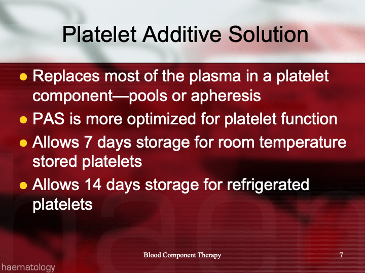
    3. Third choice:  Platelet components in platelet additive solution PAS
    4. Fourth choice:  Contact transfusion medicine for use of plasma-incompatible type
  3. Do NOT volume reduce platelets:  this adversely affects platelet function and requires a prolonged recuperation period of several hours before the platelets can be transfused.  Platelets in additive solution have only minimal residual plasma (35 ml out of 200 ml volume).
  4. There is no need to chose platelet types from a D-compatible donor because we use methods that minimize RBC contamination (automated component processing to prepare buffy coats for pooling or apheresis).
    1. It is at the discretion of the ordering physician if he wants to consider Rh immunoprophylaxis if D-incompatible type is chosen.

References:

  1. Standards for Blood Banks and Transfusion Services, Current Edition, AABB, Bethesda, MD, USA
  2. Guidelines to the Preparation, Use, and Quality Assurance of Blood Components, European Committee (Partial Agreement) on Blood Transfusion (CD-P-TS), Current Edition

Opinion: Handling Incorrect Physician Orders

Most of the non-transfusion-medicine physicians with whom I have worked have had only limited training in placing component or hospital blood bank orders.  In previous posts I have discussed this and suggested that all physicians who may possibly order blood bank tests or blood components should have a training and documented competence on a periodic basis.  Only a very few physicians, mainly hematologists and some organ transplant physicians, have placed reliable orders.

Before we had a blood bank computer system, we received orders on a manual paper requisition.  If there were errors, my technical staff and I corrected the order.  Any changes were made by me in my capacity as the blood bank medical director.  The ordering physicians and I had good relations and they had no problem with this—in fact, many were afraid of the blood bank and felt happy to be relieved of the responsibility.

In the current software era, what happens depends on how the order is placed.  Can the technical staff and I correct the order directly or must we each and every time contact the physician to revise his/her order?  Do my staff have to cancel each erroneous order and wait for the corrected order?  This could slow down the work process and prevent release of blood components in a timely manner.

Should the non-blood bank physician be allowed to order our complicated esoteric transfusion tests directly—e.g. ordering extended antigen typings or antibody identifications or elutions?   What if they order something unnecessary or inappropriate?

At a recent hospital position as Division Head of Transfusion Medicine, I discovered that the Hospital Information System HIS was very slow for all blood bank orders.  The physicians complained, I would allow them to bypass the HIS and use the manual backup system in critical situations.  If we had been forced to cancel the order and have the physician reorder, this would have greatly disrupted the work process.

We did not use the HIS at all in Transfusion Medicine.  We had patient and donor modules in the dedicated blood bank system Medinfo.  We had a limited ordering interface from the HIS to Medinfo for blood components and some basic testing (e.g. ABO/D typing, DAT, type and screen/group and save, cord blood testing, and transfusion reaction workups).  All work was done in the dedicated blood bank system.

All components were leukodepleted to < 1E6, all platelet and plasma were Mirasol pathogen-inactivated, all platelets were in platelet additive solution PAS.

The doctors did not order the specific blood component, rather they indicated a preference, e.g.

  • Packed RBCs
  • Platelets (adult dose of 2.4 E11)
  • Plasma
  • Cryoprecipitate

The following appeared as an order comment in the blood bank system:

  • The specific number or amount of the each component type
  • Preferences for pooled vs. apheresis platelets, washed RBCs, irradiated RBCs.

The blood bank staff could review the doctor’s request and order in Medinfo as per our internal protocols under my responsibility.  In effect, I was modifying the orders when needed just as I had done under our manual system.  This included type, modification, and quantity.  Yet now, I did not have to change any orders since the doctors had only indicated preferences.

For blood bank testing orders, we had our own internal algorithms for the workups.  The doctors could not order antibody identifications, elutions, or any antigen typings other than ABO/D.  They latter were triggered by the screening test results.

This system allowed us to avoid cancellations due to physician errors.  I was very comfortable taking the responsibility to alter the orders for best patient care.  If a physician did not agree with our algorithms, they could discuss the issue with one of the transfusion physicians, or ultimately appeal to me.  I was fortunate that there were no legal issues with this approach where I practiced.

In summary, my recommendations are:

  1. Have the doctors order a preference for the type of blood component.  They still order the quantity and can request modifications such as washing or irradiating or apheresis-derived or buffy coat pool platelets.  The actual allocation is made by blood bank staff under my direction.
  2. Offer algorithmic based testing.  Doctors only order basic testing which triggers reflex algorithms.  Most of the test menu is not orderable directly by the physicians (example:  an antibody identification is triggered by a non-negative antibody screen.

9/12/20

Anti-CD47 Interference

Background:

Anti-CD47 (Hu5F9-G4)interferes with all phases of pretransfusion testing, including ABO reverse typing.

Anti-CD47 (Hu5F9-G4) is a human monoclonal IgG4 antibody used to treat hematologic or solid malignancies.  CD47 is a glycoprotein expressed on all cells. 

All plasma samples reacted 3-4+ in all phases with RBCs, including immediate spin.  Stronger reactivity may occur in D-negative RBCs.  Plasma reacted with DTT, trypsin, papain, alpha-chymotrypsin or WARM (warm autoantibody removal medium—Immucor). 

DAT and autocontrol are negative or weak whereas eluates are 3+ reactive.  Reactivity is removed by multiple alloadsorptions with papain-treated cells or pooled platelets.  PEG absorption is invalid due to precipitation of the antibody.  Gamma heavy-chain specific AHG reagent (Gamma-clone IgG—Immucor) does not detect IgG4 antibodies.

Process:

  1. Always check medication history for anti-CD47 treatment.
  2. If positive, use gamma heavy-chain specific AHG reagent (Gamma-clone IgG) for IAT.

The following table summarizes anti-CD47 reactivity and compares it to anti-CD38—see reference below.

Reference:

Randall Velliquette et al, Anti-CD47 Interference in Red Cell and Platelet Testing, Transfusion 2019; 59, 730-737