The purpose of antibody titration is to determine how to follow a pregnancy at risk for hemolytic disease of the fetus and newborn (HDFN) or for organ transplant (e.g. ABO-incompatible renal or stem cell transplantation).
Titers are notoriously hard to compare across different institutions and different reagents and are subject to variation between technologists. Automated methodologies may help limit this variability.
Policy:
Indications
Pregnant patients with active anti-D to determine if a more aggressive intervention, e.g. percutaneous umbilical blood sampling, is needed to further assess the state of the fetus
ABO-incompatible renal transplantation: to determine candidacy and follow the effect of ABO column immunoadsorption and immunosuppression
ABO-incompatible stem cell transplantation to follow the course of treatment
External proficiency surveys
Procedure and Interpretation:
Titers may be done in saline or at antiglobulin phase.
Titers may be performed manually or on automated equipment (e.g. Ortho Vision Max).
If the titers are to be used as part of an external protocol, the method should be correlated with the external institution and found to be acceptable.
Titers for organ transplant (e.g. kidney) will be done at saline and antiglobulin phase.
The titer FOR PATIENT REPORTING is defined as the final tube showing a 1+ reaction.
The titer for CAP surveys will be according to the instructions of the CAP, even it does not agree with our 1+ reaction rule.
Any variances from this policy must be specifically approved by a blood bank consultant.
References:
Technical Manual, Current Edition, Bethesda, MD, USA
Standards for Blood Banks and Transfusion Services Current Edition, AABB, Bethesda, MD, USA
As our hospital network expanded, there were many patients who moved between locations. They might first start in an emergency room and then be transferred to a specialty hospital. These locations might be served from different hospital blood banks/transfusion services. What happens if work is progress from one site when the new site receives the patient. Must the previous workup be repeated or could it be used for transfusion at the next site?
For example, the ABO typing could be performed at one site and the antibody screen at a second site, and the antibody identification at still another site. Could the results be used across the entire system?
I had multiple hospital blood banks and blood donor centers. The general and specialty laboratories had multiple sites. The hospital information system was set up so that the various tests could only be performed at specific designated sites. This posed problems as patients were moved around or if some site(s) became inoperative since the specimens then had to transported at great distances for testing. Only a few basic STAT tests were available at all sites.
It was my decision to allow all test categories at all sites, e.g. a DAT request from any site, any methodology, could be used to satisfy the order. Similarly, all donor processes were available at all donor centers (the processes could be completed at one or more sites). Different hospital blood banks had different equipment but all the test categories were the same across site—the methodologies might differ. We had at least four different DATs across our system.
The interface between the blood bank and hospital system worked as follows: In the hospital information system HIS, test orders pointed to a category of testing and any methodology for that category at any site could be used in the blood bank system for testing and reporting back to the HIS. Any test in a category from any site could be used to satisfy the test request. Blood bank staff would choose the particular test methodology to use. It was NOT specified by the HIS!
In summary, for blood banks and donor centers within our system, the work could be flexibly moved between sites. There was no need to repeat testing when a patient transferred to a new site. The only type the work was repeated if testing was done at an institution outside our system.
Historically, the Lui-Freeze-Thaw elution method was used to detect ABO antibodies in suspected cases of ABO hemolytic disease of the fetus/newborn HDFN. However, the detection of such antibodies does not mean that they are clinically significant. If a clinically significant antibody is suspected, perform acid elution instead. If you want to detect ABO antibodies in a neonate or in a transplant setting, you can use this eluate against reagent A and B cells.
Policy:
For suspected cases of significant ABO antibodies (HDFN, organ transplant), perform acid-elution:
For ABO antibodies, test the eluate against reagent A and B cells using antiglobulin phase.
If the mother is ABO-incompatible with the neonate and the neonate’s DAT is positive with a negative eluate against panel cells, then test against A and B cells to rule out ABO antibodies
The same applies to organ transplant cases to detect ABO antibodies.
For detection of non-ABO antibodies, test the eluate against an antibody panel (i.e. group O cells).
References:
Technical Manual, Current Edition, AABB, Bethesda, MD, USA
Standards for Blood Banks and Transfusion Services, Current Edition, AABB, Bethesda, MD, USA
Guidelines to the Preparation, Use, and Quality Assurance of Blood Components, European Committee (Partial Agreement) on Blood Transfusion (CD-P-TS), Current Edition
In a previous post, I discussed transfusion training for hematology fellows and general pathology residents. I have no expectations that most of them have any interest in the field so I suggested concentrating on the interpretation of the direct antiglobulin test DAT and turn-around-times for services.
In contrast, the transfusion medicine physician in-training needs to understand in detail all processes, donor and patient—especially test interpretation so that he/she can make medical decisions and variances.
During my training, I was fortunate to be in a residency training program that also had an American Specialist-in-Blood Bank SBB training program. To a large extent, I attended the SBB program and even worked on the “wet” specimens.
I had no delusions that I would ever function as technologist or SBB in the blood bank. However, that extended blood bank training has made me the physician I am. I can correlate advanced, even reference, procedures to my medical knowledge and thus provide a unique offering. In contrast, even the SBB is not a physician and cannot make the medical correlations. Recently, I was flattered at an AABB meeting when the speaker thought that I was an SBB.
In certain regions where reference immunohematology laboratories and SBBs or equivalent are rare, the transfusion medicine should have sufficient technical background to help fill this gap. In my practice, I review all antibody and DAT workups and make interpretative comments for the physicians and nursing staff. These comments are entered into the blood bank computer system.
I personally tutor the trainees and make certain that they understand potentially dangerous patterns such as antibodies to high-incidence antigens, significance of the autocontrol in panreactivity, and assessing for fatal acute transfusion reactions—both hemolytic and non-hemolytic.
It also helps when I can discuss with my technical staff my interpretations and choices for clinical management. They get a better idea how important their work is for patient care and understand how any errors may adversely affect the patient.
In regions where there are good immunohematology reference laboratories, some of this may be less necessary. I lament that transfusion medicine physicians may not maintain these skills and must rely on others to their detriment. Even if one is comfortable with this, the physician is still ultimately responsible for making the clinical decision.
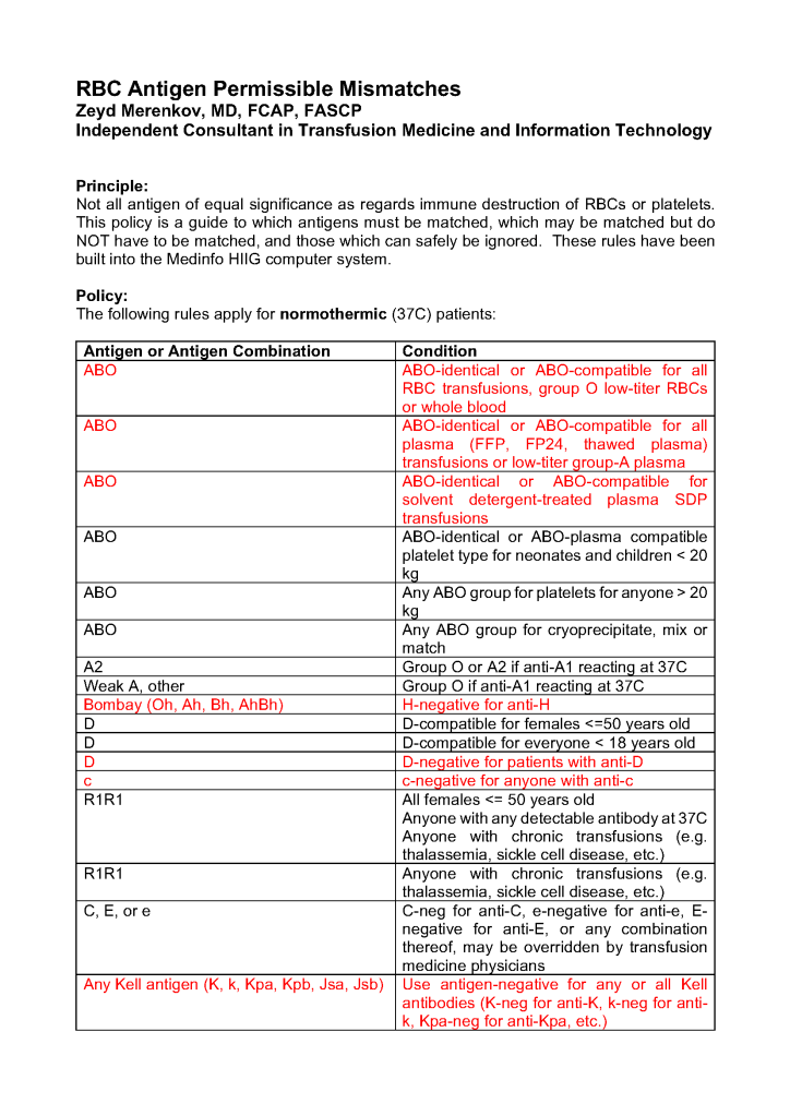
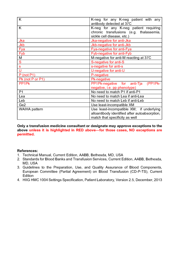
The following was my interim policy at HMC for permissible matching and mismatching. This assumes that the patient is not being transfused while in a hypothermic state.
The prohibited options were blocked in the Medinfo Hematos IIG computer system. WAIHA least-incompatible release required physician approval. This protocol did not apply to low-titer group A or low-ABO-titer whole blood units, which were not available at the time.
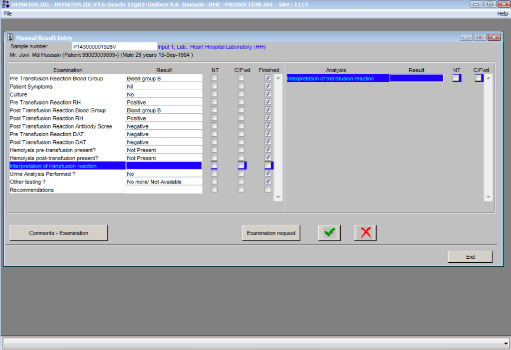
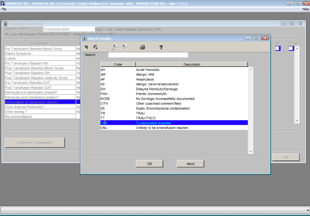
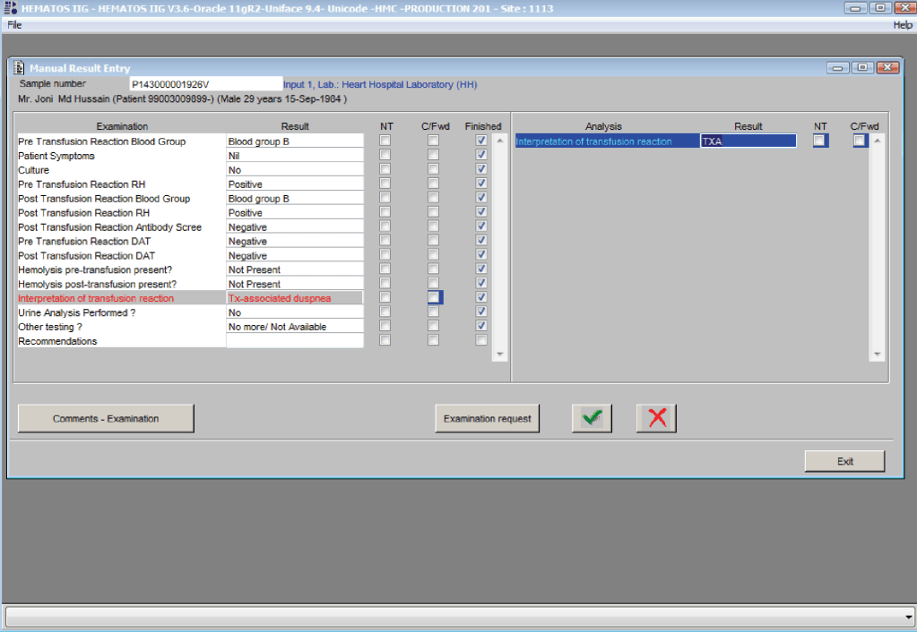
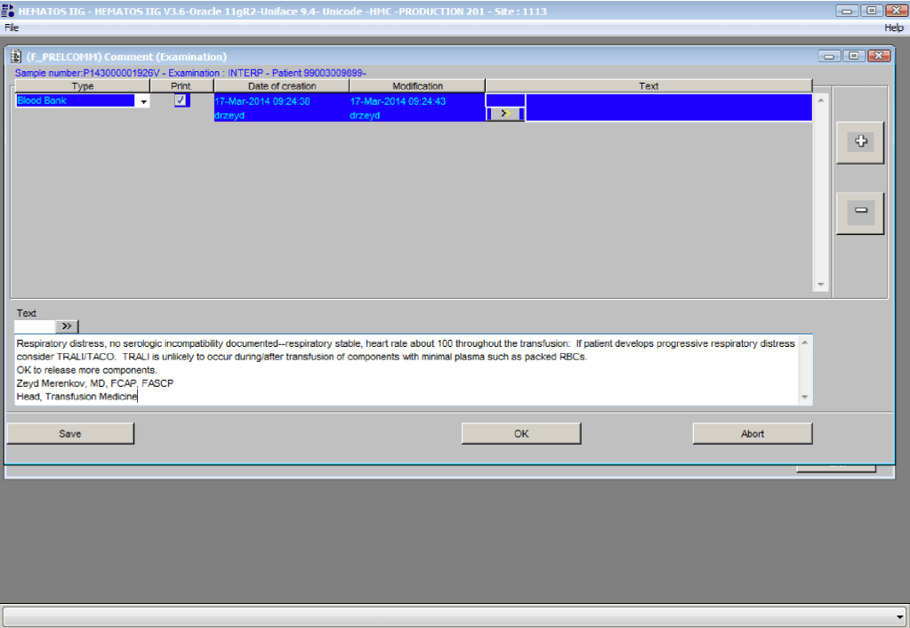
Physicians must enter their interpretations and recommendations for each transfusion reaction review. In addition, they may enter comments against any of the data. This post shows the process used in Medinfo Hematos IIG.
By highlighting Interpretation (left side), they then click on the Result Field (right side) and select their interpretation from the drop-down menu. They can also select other and then enter a comment.
References:
Standards for Blood Banks and Transfusion Services, Current Edition, AABB, Bethesda, MD, USA
Guidelines to the Preparation, Use, and Quality Assurance of Blood Components, European Committee (Partial Agreement) on Blood Transfusion (CD-P-TS), Current Edition
This policy is a reiteration of current policy to QC reagents used for antibody screening and identification to document how current practice meets these requirements. This policy is NOT a change from current practice.
Policy:
Each cell used for antibody detection must be checked each day of use for reactivity of at least one antigen using antisera of 1+ or greater avidity:
We will use reactivity encountered during the daily antibody testing: reactions of 1+ in each screening cell will be deemed acceptable. For panel cells, reactions of 1+ or greater for any specificity will be deemed acceptable.
Typing reagents such as anti-D, anti-K, anti-Fya, etc. must be checked each day of use.
Already defined explicitly in SOPs
Anti-IgG reactivity of antiglobulin reagents may be checked during antibody screening and crossmatching:
Currently performed as per manufacturer’s instructions (e.g., Immunocor, Biorad, Grifols, Ortho) for gel and tube reagents.
Typing sera and reagent cells must be checked for reactivity and specificity on each day of use, including a check against known positive and negative cells or antisera:
Already defined explicitly in SOPs
References:
Standards for Blood Banks and Transfusion Services, Current Edition, AABB, Bethesda, MD, USA
Guidelines to the Preparation, Use, and Quality Assurance of Blood Components, European Committee (Partial Agreement) on Blood Transfusion (CD-P-TS), Current Edition
Maintaining an adequate blood supply and expedited compatibility testing are critical in disaster planning. Medinfo Hematos IIG allows us to get dynamic updates of our blood supply and dynamically reallocate blood components as needed.
Policy:
Determinate total available blood supply across all locations by using the Cumulative Stock Display program in Medinfo Hematos IIG.
Recheck stock at least every hour during the disaster.
At each transfusion service site, in conjunction with a Transfusion Medicine Consultant:
Cancel reservations for elective surgical and non-emergency medical cases of affected ABO/D types.
Retain reservations for antigen-matched, oncology, NICU, and high-risk obstetrical cases.
Inform Donor Recruitment/Logistics to send SMS, radio, and television messages for blood donors—all types.
Contact ALL staff and have them report to duty.
At the Blood Donor Center, the Head Nurse, Recruitment, Supervisor, Component Processing, and Supervisor, Marker Testing will contact staff.
At hospital transfusion services, the site supervisor will contact all staff.
Process blood components using automated component technology (Reveos).
Perform all donor marker testing including single-well NAT.
Abbreviation of donor marker testing is only at the discretion of the Division Head, Transfusion Medicine.
Transfusion Services:
Release blood component according to the various protocols as needed:
Massive Transfusion Protocols
Emergency release
STAT
Priority
Routine
Compatibility testing will be electronic, immediate-spin, or full AHG as per our protocols.
References:
Standards for Blood Banks and Transfusion Services, Current Edition, AABB, Bethesda, MD, USA
Guidelines to the Preparation, Use, and Quality Assurance of Blood Components, European Committee (Partial Agreement) on Blood Transfusion (CD-P-TS), Current Edition
Although we did extended Rh and Kell typing for all cases with a non-negative antibody screen, we did not routinely do this for patients with a negative antibody screen.
Principle:
Based on recent cases of rare phenotypes in the Rh system (e.g. r’r’), we will proactively test such patients most likely for this.
Policy:
Extended Rh (C, c, E, e) and Kell typing should be done on all patients meeting the any of the following criteria:
This is the sample examination I made for the senior-most staff and candidates. This is really a projective exercise. How far can you go with this? The candidate must specify what additional information he/she needs to complete the assessment.
Name: Badge #:
Date of Exam:
Answer the following questions:
What is the ABO blood type in each of the following results:
Anti-A
Anti-B
Anti-A,B
A1 cells
B cells
O Cells
ABS
Type?
0
4+
3+
0
0
0
0
3+
0
3+
1+
4+
0
Neg
3+
1+
3+
0
3+
0
Neg
Weak
0
2+
0
4+
0
Neg
0
0
0
4+
4+
4+
Pos, all cells
Weak
0
0
4+
4+
4+
Pos, all cells
0
4+
3+
4+
2+
0
Pos, SCIII
Mf
0
0
0
4+
0
Neg
ABS = antibody screen; mf = mixed field reaction
Assume that all reactions are the same by both the tube and gel methods.
In the above table, list any discrepancies in each of the testing panels. Describe what additional tests or information, if any, are required to resolve the type.
What is ABO and D typing in each of the following results?
Anti-A
Anti-B
Anti-D
D-control
A1 cells
B cells
O cells
ABS
4+
4+
4+
3+
4+
4+
4+
Pos All cells
Anti-A
Anti-B
Anti-D
D-control
A1 cells
B cells
O cells
ABS
0
0
4+
2+
4+
4+
0
0
Anti-A
Anti-B
Anti-D
D-control
A1 cells
B cells
O cells
ABS
0
0
4+
2+
4+
4+
2+
2+
Using anti-D antisera, explain the difference in reactivity you expect between R1R1 and R2R2 cells.
Describe at least two antibody specificities associated with a mixed field reaction.
Describe the discrepancy noted in the following gel reaction and provide a differential diagnosis for the possible cause(s).
Describe when to use Diluent 1 versus Diluent 2 with the Diamed gel cards.
There is a critical staffing shortage so you are working the bench when emergency, class I blood is released (O-positive to a 25 year old male victim of a car accident). You receive the specimen and obtain the following set of results:
Anti-A—0, Anti-B—0, A1 cells—4+, B cells—4+, ABS—3+ in SCI/II/III
What actions will you take now? What blood type will you release for subsequent RBC requests? Be explicit!
An extended phenotype is ordered and Diamed Profile I-II-III cards are used. The following results are obtained on Profile Card III:
M
N
S
S
Fya
Fyb
0
0
0
0
0
0
Interpret these results.
Describe the rationale for the prophylactic use of E-c- cells in a patient only showing anti-E.
A patient develops pain at the infusion site with back pain while a unit of packed RBCs is being transfused. The following results were obtained in the subsequent workup:
Pretransfusion ABO/D: B-positive—reverse typing normal
Post-transfusion ABO/D: B-positive—reverse typing normal
Returned unit ABO/D: B-positive
Repeat crossmatch—compatible
Give possible reasons for these findings. What further investigations would you do?
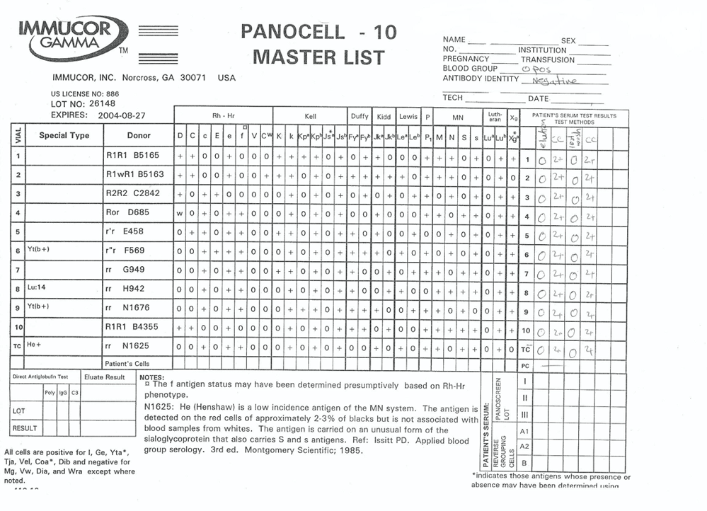
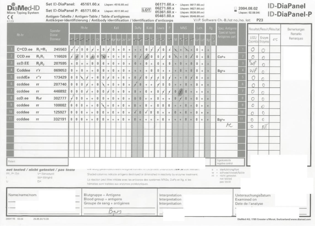
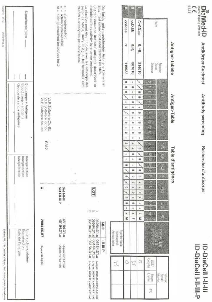
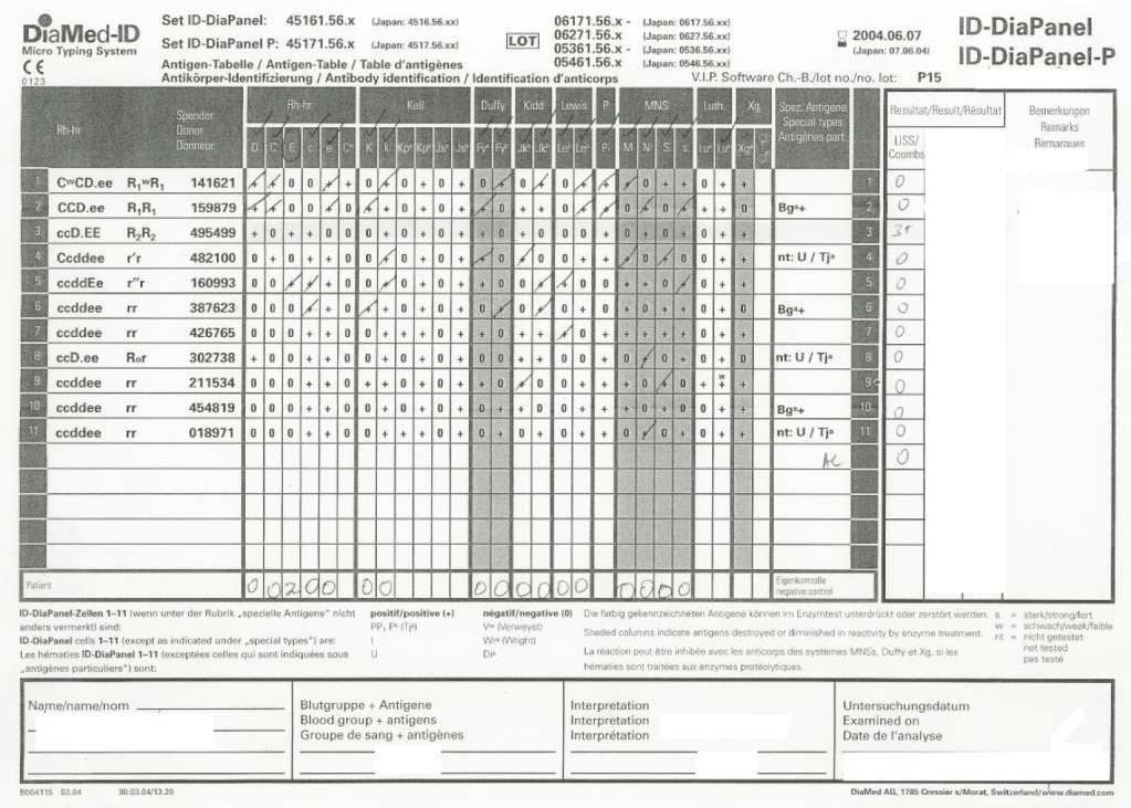
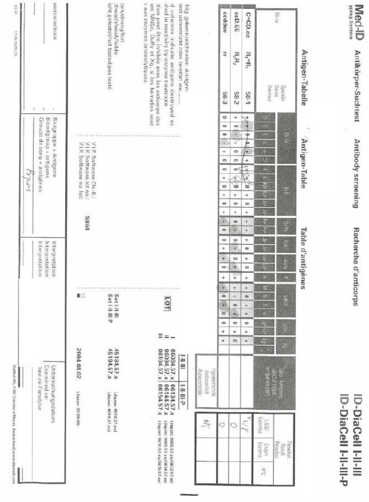
Interpret the following panels:
48 year old female with septic arthritis and severe anemia (Hgb 6.9 g/dl) and positive autocontrol 2+ (Polyspecific 2+, IgG 2+, C3d nil) and negative antibody screen:
The eluate panel results follow:
Please give your interpretation of the findings. Be sure to specify other information you may require if any.
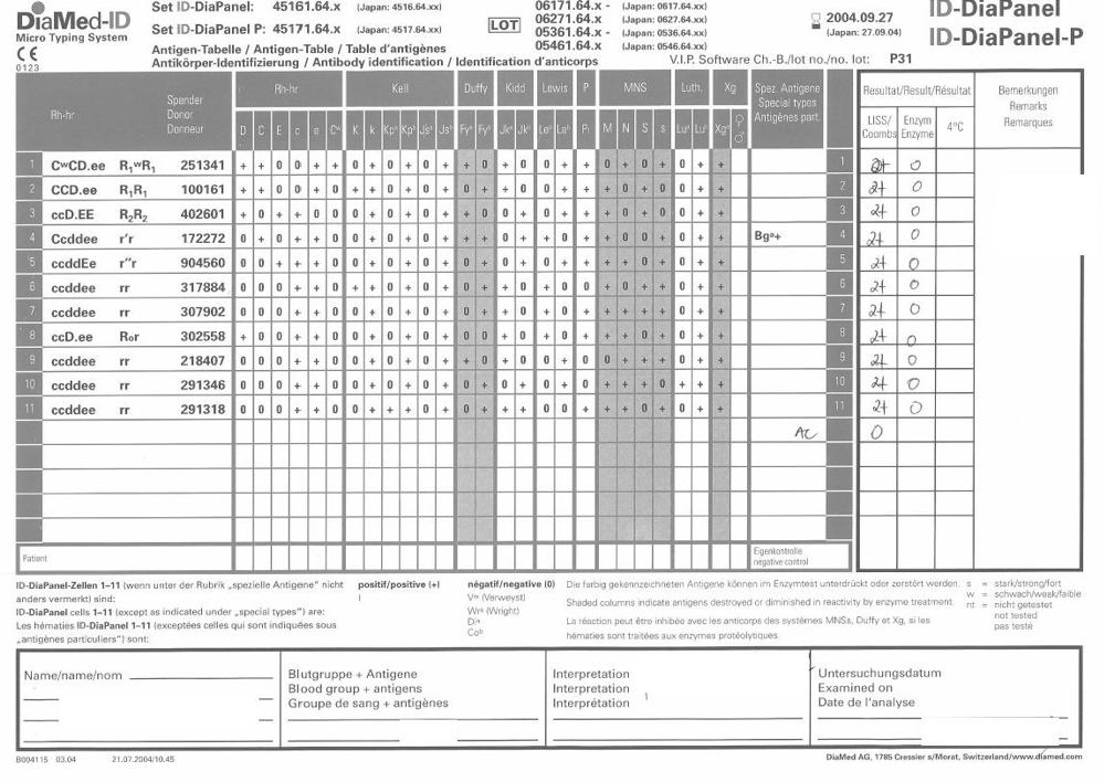
71 year old female with aortic stenosis admitted for surgical correction, blood type AB positive, antibody screen 2+ positive in all 3 cells, autocontrol negative, no history of recent transfusions.






 Dr. Zeyd Merenkov