Blocking Antibodies

This is a revised version of a previous post.

If there is strong antibody binding to an RBC, this may interfere with a typing reagent attaching to the cell and cause a false-negative, i.e. a “blocking” antibody.  Such cells may interfere with the indirect antiglobulin test IAT, i.e. the antibody screen.  The autocontrol and direct antiglobulin test DAT will be strongly positive.

The manufacturer’s instructions for using its reagents should be strictly followed in the presence of a strongly positive DAT.  If there is no reaction with the typing reagent, the result must be indeterminate.

One could try a (relatively) nondestructive elution method such as gentle-heat elution to remove some of the antibody and then retype the cells.  I have found this to be a simple and effective method for my staff to use.  Just remember that despite being “gentle,” there will still be some hemolysis present, but here it is the cells we are trying to save.

Usually, we find this situation in a neonate born of a mother with anti-D.  The baby has a strong DAT but the D typing is negative.  Check the D control carefully:  if it is positive, the result is indeterminate, try another method.  Usually gel/glass bead methods are subject to less interference.  Finally, there is always the classic saline anti-D!

In Medinfo software with a blocking antibody, a nonnegative control will trigger a manual review of the results.  There will be no automatic release.

Here is my process for handling blocking antibodies, which I set up for HMC Doha:

INTERIM POLICY:  ANTIGEN TYPINGS IN PRESENCE OF STRONGLY POSITIVE DIRECT ANTIGLOBULIN TEST (DAT):  RULE OUT BLOCKING ANTIBODY

Principle:

Antigen typing of cells with large amounts of coating antibody (i.e. strongly positive DAT 3-4+) may not always be possible because the bound antibody may block available antigen sites.  This policy is to clarify how to recognize and handle such situations.

Policy:

  1. Always follow the manufacturer’s instructions for the use of the typing reagent.
    1. In particular, note whether a control must be run with the test (e.g. D-control, D-diluent, etc.) or if it is included in the gel or glass bead card.
      1. If a control is required, use exactly what the manufacturer recommends.
      2. DO NOT SUBSTITUTE ANYTHING ELSE AS THE CONTROL!!
  2. Interpret the reactions exactly as the manufacturer indicates.
  3. If the test is invalid because of the control or any other reason, report the antigen typing as indeterminate and send for Transfusion Medicine Physician TMP review.
  4. If the DAT is 3-4+ and the antigen typing shows no reaction (apparent negative), send the case to the Transfusion Medicine Physician for review and final interpretation.  DO NOT ENTER THE RESULT AS NEGATIVE UNLESS THE TMP INSTRUCTS YOU TO DO THIS!!
  5. To rule out a blocking antibody, a special elution to gently remove the coating antibody may be needed so that the RBCs can then be typed (not acid glycine technique—rather use gentle heat elution.)  The Transfusion Medicine Physician will decide whether to do this additional testing.

References:

  1. Standards for Blood Banks and Transfusion Services, Current Edition, AABB, Bethesda, MD, USA
  2. Technical Manual, Current Edition, AABB, Bethesda, MD, USA
  3. Guidelines to the Preparation, Use, and Quality Assurance of Blood Components, European Committee (Partial Agreement) on Blood Transfusion (CD-P-TS), Current Edition

Overextending Oneself—Two Case Studies in the Blood Bank

One has to learn when enough is enough.  There are times when there are staff shortages but the conscientious staff wants to be the Super-Tech and handle all the work, whether or not there are sufficient resources.  This is a big gamble, and there may be serious consequences for the over-achiever and for the patient.

Anecdote #1:  Chicago Blizzard of 1979 (13-14 January):

When I was in my residency training in Chicago, I was in the blood bank during the blizzard of January, 1979.  The following tragedy occurred.

Suse was one of the best blood bank technologists that I have ever known, extremely conscientious and very meticulous—and very fast at doing things.  She was a workaholic.  Suse’s whole life centered on her job at our academic medical center—so much so that she had an apartment near the hospital complex.  In mid-January, a snow storm was predicted with an estimated snowfall of about 5 cm. total.  Actually, that night a blizzard developed and around a meter of snow fell with white-out conditions and zero visibility.  Preoperative patients had been admitted the night before based on the low snowfall prediction.

The next day was chaos.  Essentially only staff who lived near the hospital complex could report to work.  Suse came in and saw all the pending preoperative blood requests.  She decided to “double-up” and work on two cases at one time.  In the rush, she mixed up test tubes and issued ABO-incompatible blood for a surgical case.  The surgeon noted the abnormal oozing of blood at the operative site and stopped the transfusion.  Hematuria developed, but the patient survived.

Suse was suspended pending investigation.  Based on her excellent work record, she was offered to return to work.  Unfortunately, she became very depressed and was afraid to return since she feared she would make another mistake.  She never worked in the blood bank again.

Anecdote #2:  Shortage of Blood at Major Hospital:

1991 in another country, a large hospital complex was suffering a shortage of blood.  A large number of donors were called and the available staff were overwhelmed with work.  One donor phlebotomist decided to collect whole blood from two different donors simultaneously and in the confusion, mixed up the sample tubes for donor marker testing.

Unfortunately, one of those donors was HBsAg positive, but with the specimen mix-up was marked as negative.  The unit of blood was transfused, and the recipient developed fulminant hepatitis B and died.

Analysis:

In both these systems, there were processes in effect not to work on two patient specimens or collect two donors at one time, but the staff took short-cuts.

No one is super-human.  Don’t try to cut corners and handle more than one patient at a time.  Your intention may be good, but you will be judged by the consequences.  No one will care about the extenuating circumstances.  You will be blamed.  I tell my staff that if they cannot handle the workload, they should contact me as the Division Head, Transfusion Medicine, to triage the cases for them.  My role is to bring these events to the higher authorities to get the resources we need to do the work properly and safely.

Case Report: Overwashing During Elution

Note:  This is a repost.

I cannot emphasize enough proper technique in doing the washing during the elution process.  We are usually concerned about too little washing and thus possibly residual reactions in the last wash.  However, aggressive overwashing may remove the bound antibody resulting in a negative result.

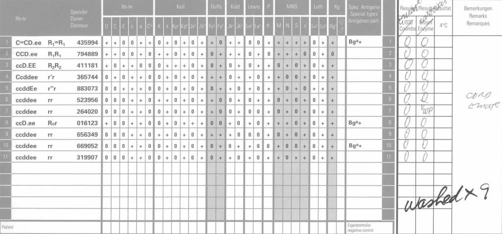
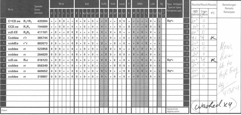
Here is an example of anti-PP1Pk (alias anti-Tja).  The mother’s panel shows an antibody to a high prevalence/incidence antigen with negative autocontrol and no lability at enzyme phase:

The neonate’s DAT was weak positive at polyspecific and IgG monospecific phases.  An eluate was performed.  Here is the result after washing four (4) times:

Since 2 cells in the last wash were very weakly positive, the washing was continued for a total of 9 times with the following results:

Even then there was very weak positivity in one cell, but the eluate was negative.  We had washed away the attached antibodies.

My Opinion: Use of Enzyme Panels

This is an updated version of a previous post.

Working for many years in the Middle East/Gulf, I have encountered significant antibodies that can only be detected at enzyme phase.  This is especially true of Rh system antibodies, particularly anti-c in an R1R1 patient.  I have attached an example.

The reasons I strongly recommend this practice are:

  1. Weak Rh system antibodies (as above)
  2. Confirmation of enzyme-labile antibodies, especially if there may be combinations of enzyme-labile and enzyme-resistant antibodies (e.g. anti-Fya and anti-c).

It is also important to consider which enzyme to use:  bromelin, ficin, or papain usually and sometimes trypsin or chymotrypsin.  They do not always attack at the same site.

In addition to most common MNSs and Duffy system antibodies, many Kell antibodies (e.g. K or K1, Kpa) are labile with papain:  however, with ficin they may be partially labile, unaffected, or even enhanced.

Using enzymes is a double-edged sword since they may enhance cold antibodies and thus cause nonspecific reactions.  Thus, I know many of you may not routinely include them in your workups.

It is essential to follow the manufacturer’s recommendations for their use.  If you make your own enzyme-treated cells and prolong the incubation, you may get false positivity.  You should also be careful about using potentiators with enzyme-treated cells—normally I run them in saline.

Since anti-c may cause severe hemolysis and severe hemolytic disease of the newborn, I am especially vigilant in my R1R1 patients, particularly females of child-bearing age and all chronically transfused patients.  I prophylactically match R1R1 patients with R1R1 RBCs in these categories, regardless if either anti-E or anti-c are expressed.  I have seen many examples where the anti-c is only detected at enzyme phase.

It is my practice to always include an enzyme panel.  I would be very interested to know your practices?  When do you use enzymes?

Order of the Steps in Serologic Testing

This is a teaching document I give to new staff, medical technology students, pathology and residents.  Very often I get the question, “Why can’t I just do the antiglobulin phase crossmatch first and then phenotype the RBC unit?” Or:  “Why do I have to add reagents in a particular order?”

My practice has always been to select an antigen-negative RBC unit first, then do the antiglobulin-phase AHG crossmatch.  This way I know that the unit was definitely phenotyped before release.  Likewise, the blood bank computer now only offers antigen-negative units for allocation and then crossmatching if there is a clinically significant antibody.

In a manual setting without a blood bank computer system, performing the AHG crossmatch may yield a negative result, even if the unit is antigen-positive.  With storage, some antigenic expression is weakened so it may not be detected at the time of crossmatch.  Yet, there may still be enough antigen present to cause hemolysis.  Not detected does not necessarily mean not present!!

I expect that many inexperienced staff may be tempted to forego the antigen typing if the AHG crossmatch is negative.

This is an analogous logic to the question, “Do I add the cells or the antiserum/plasma/serum first for the reaction?”  If you add the cells first, you may forget to add the patient’s plasma/serum or a typing antiserum and you might not be able to detect the omission by looking at the tube or gel.  Actually, I once recommended to one vendor that it color the typing antiserum so it was conspicuously showing on the gel.

I was taught that this is a matter of discipline to ensure that all steps are performed.

However, for every practice, there has to be flexibility.  If there is no typing reagent or if it is very expensive or in short supply, one may have no choice but to do the AHG crossmatching first.  Often there is still another option:  one can often preliminarily screen units first before using a rare reagent—examples:

  • Check if patient is group O first and antibody screen panreactive in suspected anti-H.
  • Check P1 typing first if there is a suspected anti-PP1Pk.
  • Check the antibody screen for panreactivity first for antibodies of high-incidence or prevalence antigens.

ABO Incompatible Stem Cell Transplant: What ABO type of RBCs Should I Use?

In an ABO-incompatible stem cell transplant, both donor and recipient RBC types may be present.  Likewise, immune effector cells from both the donor and recipient may be present.

Using group O RBCs and AB plasma is an option but there are limited supplies of both.  Since we use RBCs in additive solution (SAGM), only minimal residual donor plasma is available and unlikely to be clinically significant.

Here is my approach:

  1. Use fresh specimen to perform forward and reverse type
  2. Check the reverse type:  does it show either anti-A and/or anti-?
    1. If anti-A detected, do not transfuse group A RBCs.
    2. If anti-B detected, do not transfuse group B RBCs.
    3. If both anti-A and anti-B detected, continue using only group O RBCs.