Nonspecific Reactions due to Reagents Near Expiration Date

This is an updated version of a previous post.
Whenever I had a “nonspecific” antibody, I had to first rule out issues with the reagents themselves. The following example shows weak to 2+ reactions in the panel cells and autocontrol.

The variability in the reactions made me initially uncomfortable about called this WAIHA. I then checked the panel details: the testing was done only six days before the panel outdate.

I told my staff to repeat the workup with the new panel expiring five weeks later. The difference is astounding!!

Remember:  if you work in the Middle East, the environmental conditions can be extreme in summer (>50C).  Do you know how your reagents were handling during transport?

My advice: if you are concerned there is a clinically significant antibody but cannot confirm it, consider repeating the workup using fresh reagents and a fresh specimen. Repeating the workup after a few days may make the detection clearer.

Original Panel with Near Expiry Reagents:

Test performed 7/2/06, reagents expire 13/2/06:

Same Patient with New Reagents and New Sample:

Test Performed 14/2/06, reagents expire 20/306:

Rare Jka Antibody

This is a repost of this rare antibody with a few new additional comments.

This is a rare anti-Jka antibody that only reacts with polyspecific AHG and only in homozygous Jka+ cell.  In the past three decades, I have only seen three of these.

Almost all anti-Jka and anti-Jkb antibodies can be detected using heavy-chain gamma specific AHG.  I prefer to use this reagent since it minimizes interference from cold antibodies.

Whenever you have a nonspecific antibody, never discount the possibility of a Kidd antibody.  Consider doing extended phenotyping and include Jka and Jkb typing and always check for dosage effect.  I have a specific post on my approach to nonspecific antibodies.

Select antibody screening cells including homozygous Jka+ and homozygous Jkb+.  This usually requires a three-cell screen.

Never forget that a negative result only means not-detected.  If there is other evidence for hemolysis, consider performing additional studies.

Physician Comments in Medinfo

Principle:

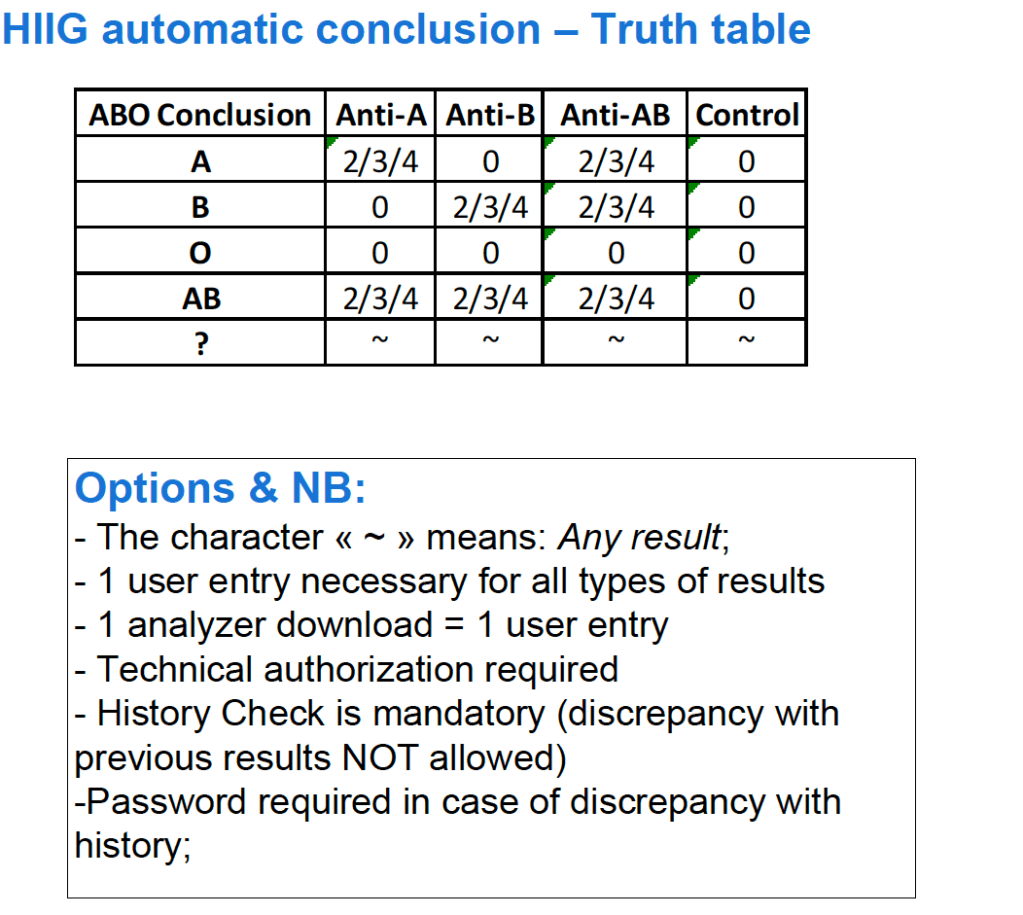
There are several different types of comments in HIIG:

  • Donor Global
  • Patient Global
  • Analytical Comments
  • Result Comments

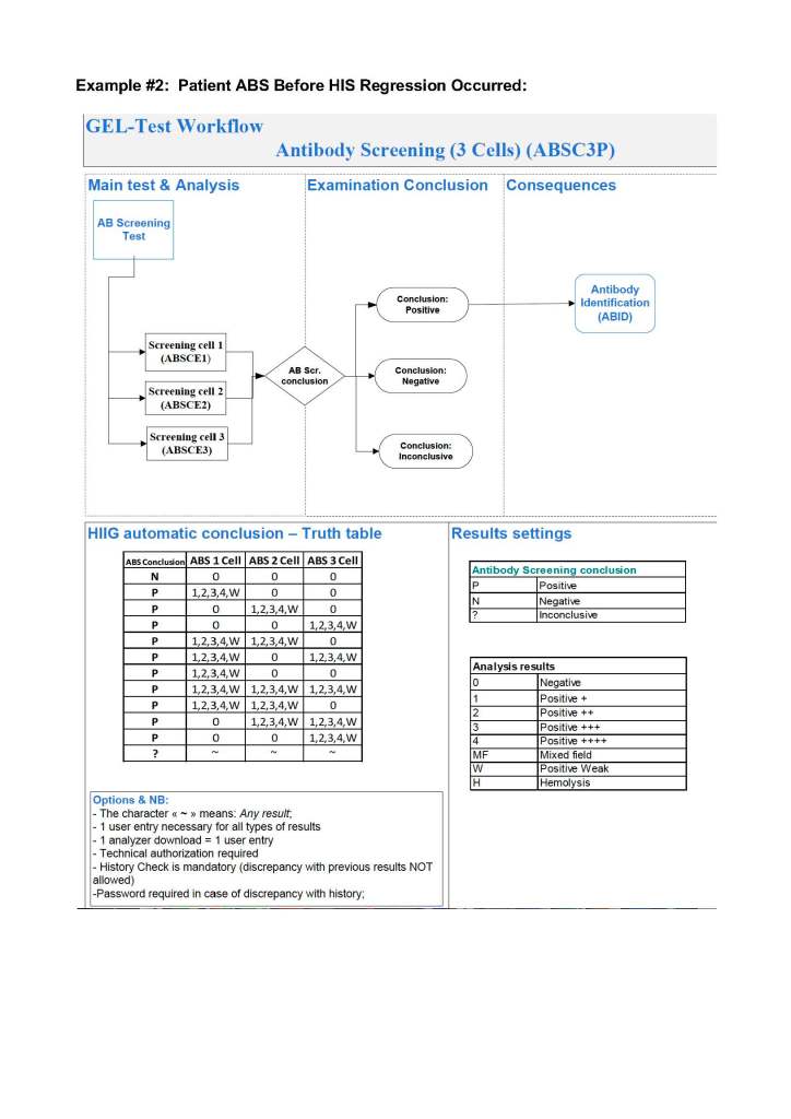
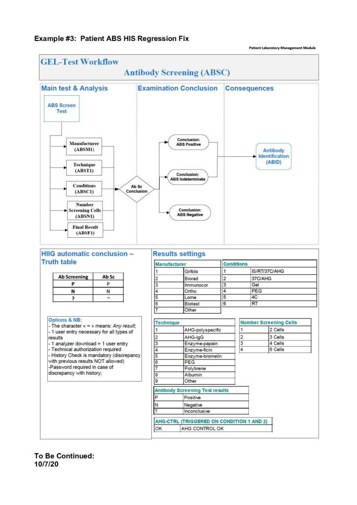
Physicians may enter any of these comment types.  Comments may be entered before or after a test is authorized/verified.  If entered after authorization, the test must be modified to accept the comment.  Only result comments are visible in the patient’s medical record.

Policy:

  1. Physicians should use comments in the following situations:
    1. Interpretations of DAT, antibody identification, ABO discrepancies
    2. Instructions for the selection of specific and/or modified blood components
    3. Donor eligibility issues (e.g. donor marker testing abnormalities and disposition)
    4. Donor reactions
    5. Telephone call documentation
    6. Donor counseling documentation
    7. Any special instructions to staff
    8. Any other situation where the transfusion physician determines it is desirable to enter a comment
  2. Copy all result-comments and also enter then as global comments against the patient or donor record as applicable.

Procedure—Patient Result Comments After Results are Authorized/Verified:

Sign into HIIG and select Patient module, Patient Consultation, and enter the patient hospital HC number.

Comments in Medinfo Hematos IIG

This post is the policy for using comments in Medinfo software.  A subsequent post will show the process of entering comments.

Principle:

There are several different types of comments in HIIG:

  • Donor Global
  • Patient Global
  • Analytical Comments
  • Result Comments
  • Contraindication Comments

Global Comments appear on the first main screen of either the donor or patient record.  The presence of comments is indicated by a bar at the bottom of the screen (in yellow or blue saying Presence of Comments.  Double-clicking opens the list of entered comments.

Examination/Results Comments appear only when you open the result to which it is attached.  You must know in advance to which result they are linked to find them.

Contraindication Comments appear when entering a donor deferral code

At HMC, we will enter examination/results comments again as global comments (donor or patient) so it is easy for staff to retrieve them and see them with all other comments.  You can do this by cut and paste.

Physicians may enter any of these comment types.  Comments may be entered before or after a test is authorized/verified.  If entered after authorization, the test must be modified to accept the comment and require a special password (not the user sign-into HIIG).  Only results/examination comments are visible in the patient’s medical record.  Global, analysis, and contraindication comments are visible only in HIIG!  Donor comments are only visible in HIIG.

The presence of comments documents physician review of abnormal results as required by the various accreditation standards.

Policy:

  1. Only designated staff may enter comments.
  2. Comments entered after authorization/verification of results will modify the donor/patient record and require a special, high-level password distinctly different from the normal user password.
  3. Enter comments in the following situations:
    1. Telephone call documentation (e.g. critical values).
    2. Interpretations of donor or patient test results, transfusion reactions, etc.
    3. Instructions for the selection of specific and/or modified blood components
    4. Donor eligibility issues (e.g. donor marker testing abnormalities and disposition)
    5. Donor reactions
    6. Donor counseling documentation (e.g. donor counseled at 0930 on 24/3/14 about his abnormal result).
    7. Any special instructions to staff
    8. Any other situation where the transfusion physician/supervisor or designate determines it is desirable to enter a comment
  4. Copy all results/examination-comments and also enter then as global comments against the patient or donor record as applicable.

References:

  1. Standards for Blood Banks and Transfusion Services, Current Edition, AABB, Bethesda, MD, USA
  2. Workflows 1001-1005, Medinfo Hematos IIG, 2013-2014.

Blood Component Transport Temperature Monitoring

Principle:

Blood components must be maintained at specified temperatures to avoid hemolysis, bacterial contamination, and maintain full efficacy (e.g. coagulation factor activity.)

Policy:

  1. Freshly collected whole blood for blood component preparation must be kept between 20-24C if  platelets are to be made.  Otherwise, it must be maintained between 1 and 10 C.
  2. Prepared RBCs and thawed plasma must be transported between 1 and 10 C.
  3. Platelets (pools and apheresis), thawed cryoprecipitate, and granulocyte concentrate must be transported at 20-24 C.
  4. Frozen components (frozen RBCs, FFP, FP24, cryoprecipitate) must be kept frozen during transport.
  5. There must be an appropriate means of documenting that the proper temperature was maintained.  Examples of compliance may include:
    1. LCD stickers that change color if the component goes outside the selected temperature range.
    2. Digital temperature recording systems—Examples:
      1. TempTale ® or other temperature recording devices (added to a transport container without a temperature-controlled container)
      2. Temperature controlled transport containers with integral recording systems
      3. The recording session for each transport episode should be downloaded, reviewed and saved.
  6. If the temperature goes outside the specified temperature range, the components cannot be used for transfusion or manufacture.
  7. All devices must be validated to meet their specified temperature-recording capabilities before being used.
  8. Specific SOPs for the use of the selected monitoring devices must be prepared and in use.

Note the different temperature ranges for refrigerated components stored in the blood bank (1-6C) versus transport (1-10C).

References:

Section 5.6.5, Standards for Blood Banks and Transfusion Services, Current Edition, AABB, Bethesda, MD, USA

DAT-Positive Hemolytic Anemias

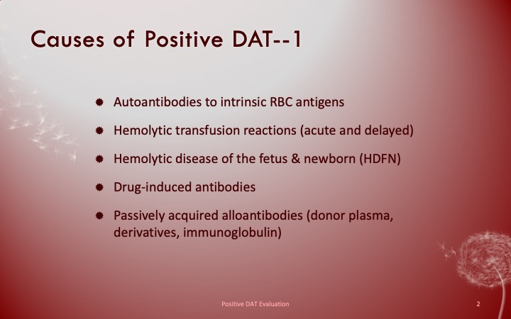
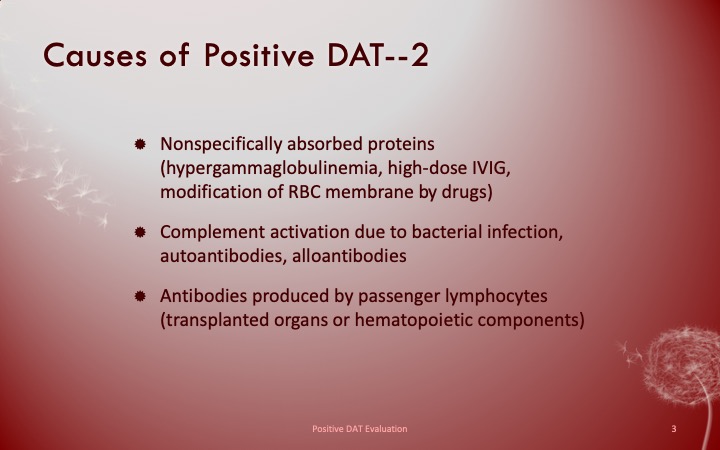
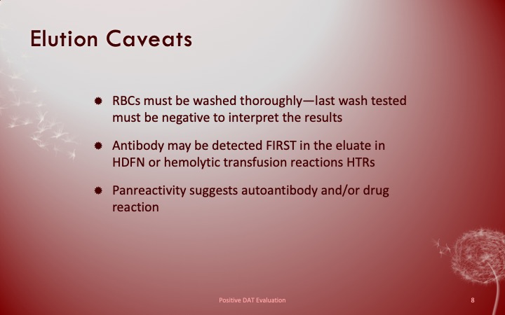
This is a helpful Medscape diagram showing causes of direct-antiglobulin-test-positive hemolytic anemias.  Note that not all cases of the conditions may consistently be DAT-positive, especially the ones that fix complement.  Again, a negative result means non-detected, not necessarily not present.

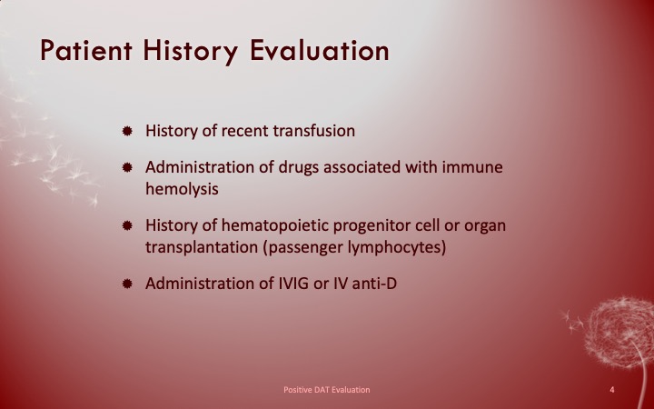
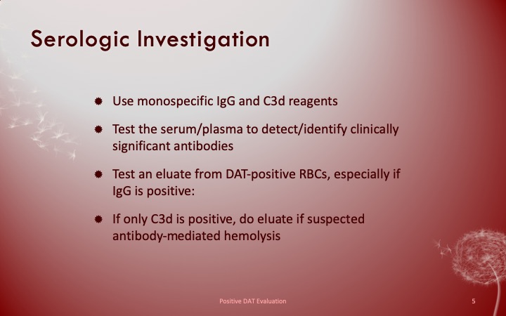
In my own test algorithm, if hemolysis is suspected, I would perform monospecific IgG and C3 DATs.  I might also include a special DAT card detecting both C3c and C3d and heavy-chains mu (IgM) and alpha (IgA) if the initial DAT is negative.  C3c positivity would tell me that active complement activation is occurring.

Handling Nonspecific Antibody Panel Reactions

Note: this is an updated version of a previous post.
Anyone reviewing antibody panels, especially in the Middle East/Gulf region, encounters many panels for which no antibody specificity is identified.  As a transfusion medicine physician who often got called during the night for release of RBCs for patients with “nonspecific” pattern, this was a big headache.

Is it “nonspecific” because there isn’t a clinically significant antibody OR the technologist did not perform the testing or its interpretation correctly?  Does it need further testing?  Do I release blood components at this time?

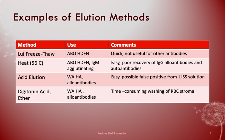
In general, I do not routinely use polyspecific AHG for routine testing.  My first choice is for a gamma heavy-chain specific AHG but this is not available for gels or glass beads.  Then, I select the IgG AHG even though it does react with light chains and can detect IgM cold antibody reactions.

In general, with nonspecific reactions, I recommend the following:

  1. Repeat with a gamma heavy-chain specific reagent if the initial workup was made with another type of AHG (polyspecific or whole molecule IgG).
  2. Always do enzyme panels, sometimes with both papain and ficin reagents:  many Rh antibodies are optimally detected only at enzyme (example:  R1R1 patient with only anti-E at AHG phase but anti-E and anti-c at enzyme).
  3. Perform an extended Rh/Kell/Duffy/Kidd/Kell/MNSs/P1 phenotype and specifically check for those negative typing results AND for dosage (could the antibody only be detected in homozygous cells like many anti-M are?).
  4. Perform classical room temperature, 37C, and finally AHG phase testing.  Routinely I do not do this since antibodies not detected at 37C are unlikely to be clinically significant.  Sometimes, the AHG phase reactivity is a cold antibody of high-thermal amplitude.
  5. If the Jka or Jkb antigen typings are negative, repeat using a polyspecific AHG reagent.
  6. Use additional panels from multiple manufacturers.  Some reagents detect more nonspecific reactions than others.
  7. Try other potentiators than LISS such as PEG.
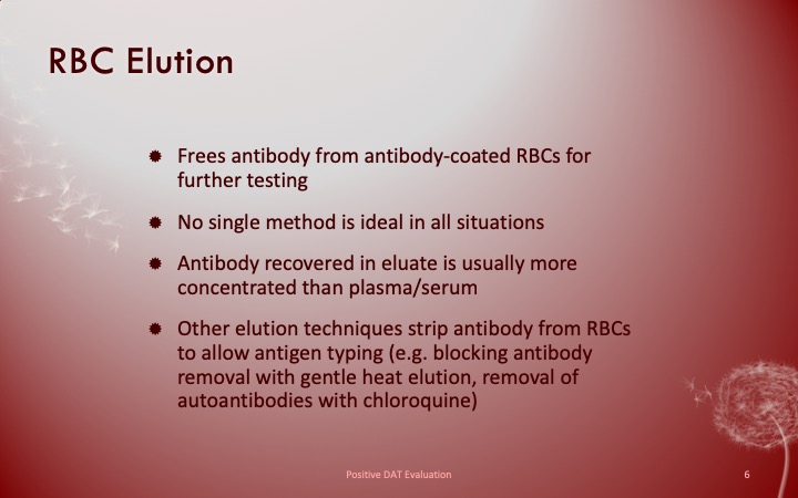
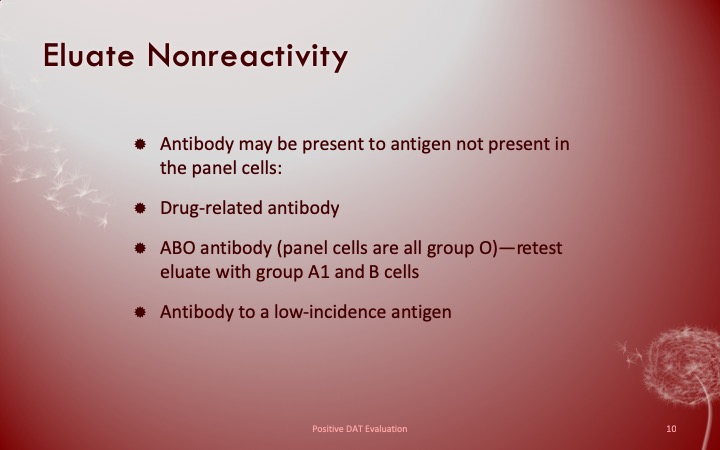
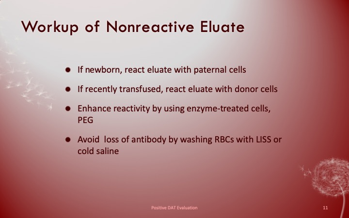
  8. Check the outdate of the panel and reagents:  if less than 1 week remaining, consider repeating with fresh reagents and getting a new patient sample.

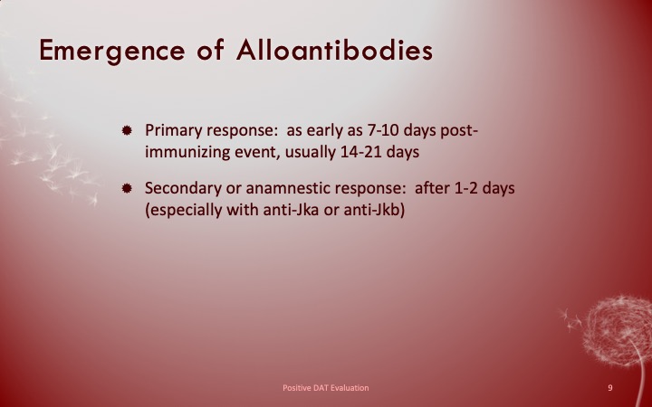
Finally, if you still cannot define the specificity, consider repeating the testing after several days.  Maybe it is a newly emerging or an anamnestic response.

I emphasize as a physician, I do not care to see all possible antibodies present in the specimen but rather only those likely to be clinically significant.  In general, there is a shortage of labor in the blood banks so I want to eliminate unnecessary work.

Autologous Transfusion

This a revised version of a previous post for the processes of autologous transfusion that I developed at HMC Doha.  It can serve as a template for other sites and was also a teaching document for the Transfusion Committee members.

Background:

There are four basic types of autologous transfusion:  preoperative, perioperative hemodilution, intraoperative, and postoperative drainage/collection.  The use of all of the above techniques can significantly decrease the need for homologous blood and as an added benefit reduce the risk of the disease transmission and immunosuppressive effects of such homologous transfusions.

Preoperative collection can make available packed red blood cells, whole blood, platelets, FFP, and/or cryoprecipitate.  However, at most two units of blood per week can be collected.  RBC’s can be stored for up to 42 days in the liquid state, frozen RBC’s up to ten years, platelets up to five days, and fresh frozen plasma and cryoprecipitate up to one year.  The last collection cannot be less than 72 hours prior to the surgery time.  Units can be collected as long as the patient’s hematocrit remains above 33%.  Supplemental iron and erythropoietin can increase the number of units harvested.  The biggest obstacle to using this service is the coordination of the patient scheduling for this procedure.  The blood bank does not have the resources to prospectively analyze the surgical scheduling and make the various appointments, contact the attending physician, etc.  Thus, this service is vastly underutilized.

PHD or Perioperative hemodilution (also called acute normovolemic hemodilution) is useful in cases when the anticipated blood loss is at least one liter and the initial hematocrit is at least 34%.  This includes essentially all types of surgery, but in particular cardiac, vascular, orthopedic, and urologic cases.  The patient’s hematocrit Hct. is lowered to the range of 20-25% and the blood is replaced by crystalloid in a ratio of 3:1–i.e. three times as much fluid as blood, or in the case of colloid replacement, a 1:1 ratio of colloid plus 0.5 to 1.0 ml. of crystalloid.  Crystalloid has the advantage of being readily removed by diuretic use.  However, this technique should not be undertaken when vascular access is inadequate or appropriate monitoring devices are lacking.  The physician performing PHD must be familiar with the compensatory mechanisms normally invoked when the hemoglobin is acutely lowered.

Another new twist to PHD is the perioperative collection of platelets by a special attachment to a cell-saving machine.  This could allow collection of a typical apheresis load, about 6 to 10 units of fresh platelets for potential use.  There are currently studies underway to determine if this has particular clinical advantages to warrant the additional cost.

Intraoperative salvage may be performed with a number of canister or automated devices.  The latter is usually used when there are large volumes (usually 3 or more units) of blood to be salvaged.  Depending on the body site, the recovered material is at least filtered and may or may not be washed.  Care must be taken to collect the blood at a low suction rate and with minimal turbulence to minimize hemolysis.

Postoperative drainage collection of certain sites such as post-knee replacement surgery or chest wounds involves a canister collection device.  This blood may or may not be filtered before reinfusion.

Note that perioperative and intraoperative material can only be transfused up to six or eight hours at room temperature or 24 hours if refrigerated at 1-6 degrees (depending on the method used) post collection to minimize the risk of infection.  Intraoperative collection may be contraindicated in cases of cancer and if the bowel has been violated.

Other Issues:

The transfusion criteria for autologous blood is the same as for allogeneic units.

The same compatibility testing algorithm applies both the autologous and allogeneic units.

Policy:

  1. Scope:
    1. Predeposit collection of Whole Blood/RBCs and plasma is under the authority of Transfusion Medicine.
    2. Perioperative hemodilution, intraoperative cell salvage, and postoperative drainage collection is under the authority of the National Transfusion Committee in conjunction with the Departments of Surgery and Anesthesia.
      1. The Division Head, Transfusion Medicine will liaise with the clinical departments as needed.
      2. Transfusion Medicine may provide blood collection bags for perioperative hemodilution upon request.
    3. Transfusion Medicine does not receive autologous collections—perioperative, intraoperative, or postoperative drainage collection.
  2. Processes directly under Transfusion Medicine authority:
    1. Autologous collection of whole blood/RBCs (pre-deposit) for elective surgeries may be considered especially if:
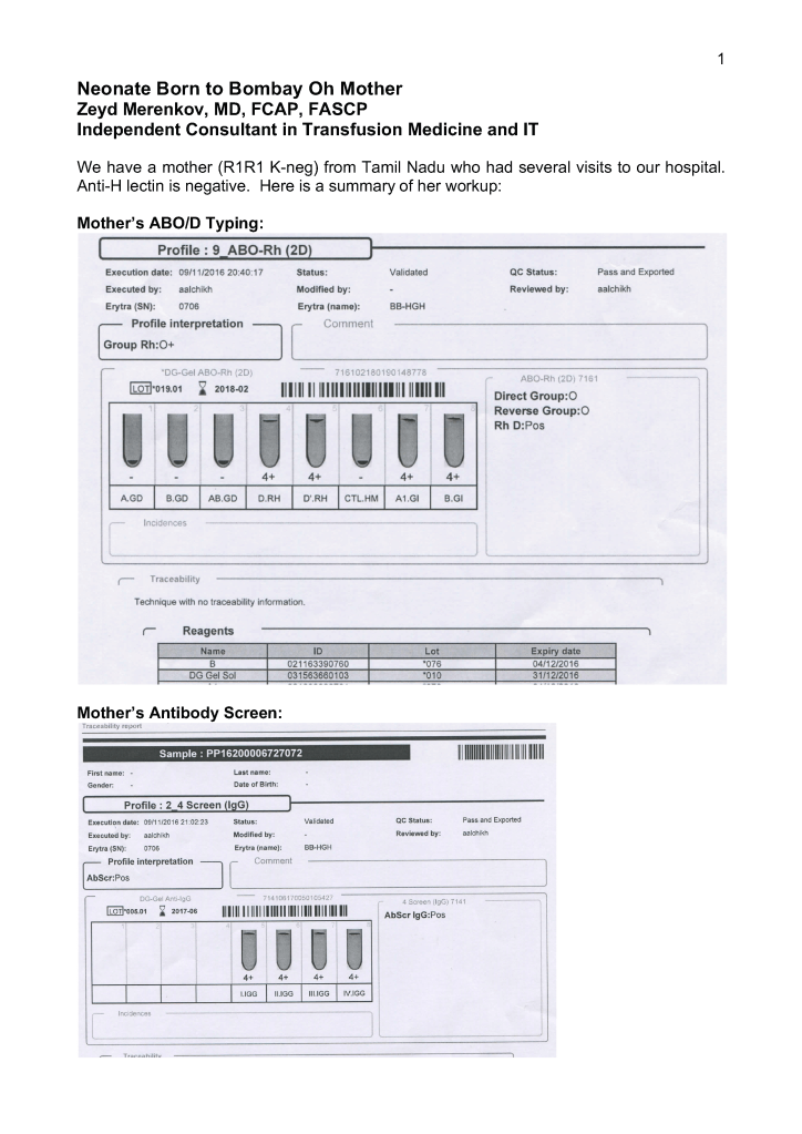
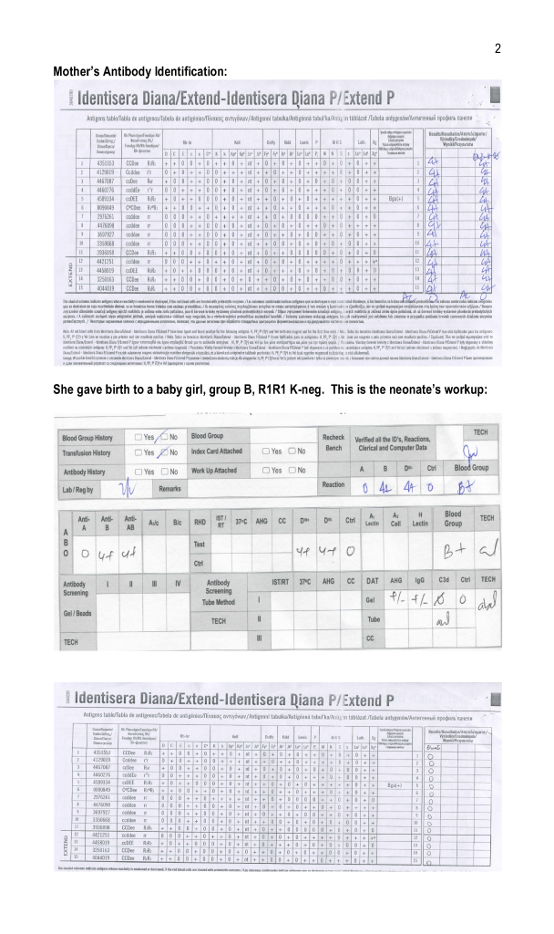
      1. The patient has a dangerous antibody for which antigen-matched units cannot be easily obtained (e.g. anti-k (cellano), anti-PP1Pk (anti-Tja), anti-H (Bombay and Para-Bombay phenotypes).
    2. Autologous collection of plasma may be considered for patients with IgA deficiency with documented specific anti-IgA antibodies.
    3. Other requests will be reviewed by the Head, Transfusion Medicine or designate.
    4. The final decision to proceed with items 2.1 and 2.2 will be made by the Head, Transfusion Medicine or his designate.
  3. Process for Transfusion Medicine Autologous Procedures
    1. The requesting physician shall provide a written or electronic order to the Blood Donor Center.
    2. The request will be reviewed by a transfusion medicine physician.
    3. If rejected, the requesting physician will be notified with the reason for the rejection.
    4. If approved, the donor shall be screened by the usual donation process except:
      1. Hgb >= 11 g/dl will be acceptable for whole blood collection.
      2. Females may also donate autologous plasma.
      3. The last autologous donation will be at least 72 hours before the elective procedure.
    5. Marker testing:  Components from autologous donors with confirmed positive cases of HBV, HCV, or HIV will NOT be used for autologous donation and will be destroyed.
    6. Computer:  Autologous collections will be entered as specifically at the time of registration in the Medinfo  Hematos IIG blood bank computer system and be labeled as autologous in their corresponding ISBT labels.
    7. The transfusion criteria for autologous units shall be the same as for homologous blood.
    8. Both autologous and allogeneic units will follow the same compatibility testing algorithm.
  4. Responsibilities:
    1. Predeposit:  Directly under the control of Transfusion Medicine for all aspects:  policies, procedures, and direct performance of the procedures, including annual review of criteria
    2. Perioperative:  Division Head, Transfusion Medicine involved in conjunction with Surgery and Anesthesia through the National Transfusion Committee.
    3. Intraoperative:  Division Head, Transfusion Medicine involved in conjunction with Surgery and Anesthesia through the National Transfusion Committee.
    4. Postoperative:  Division Head, Transfusion Medicine involved in conjunction with Surgery and Anesthesia through the National Transfusion Committee.

References:

  1. Standards for Blood Banks and Transfusion Services, Current Edition, AABB, Bethesda, MD, USA, 2014
  2. TRM.41600 CAP Checklist Standard