Autologous Transfusion

This a revised version of a previous post for the processes of autologous transfusion that I developed at HMC Doha.  It can serve as a template for other sites and was also a teaching document for the Transfusion Committee members.

Background:

There are four basic types of autologous transfusion:  preoperative, perioperative hemodilution, intraoperative, and postoperative drainage/collection.  The use of all of the above techniques can significantly decrease the need for homologous blood and as an added benefit reduce the risk of the disease transmission and immunosuppressive effects of such homologous transfusions.

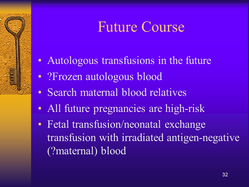
Preoperative collection can make available packed red blood cells, whole blood, platelets, FFP, and/or cryoprecipitate.  However, at most two units of blood per week can be collected.  RBC’s can be stored for up to 42 days in the liquid state, frozen RBC’s up to ten years, platelets up to five days, and fresh frozen plasma and cryoprecipitate up to one year.  The last collection cannot be less than 72 hours prior to the surgery time.  Units can be collected as long as the patient’s hematocrit remains above 33%.  Supplemental iron and erythropoietin can increase the number of units harvested.  The biggest obstacle to using this service is the coordination of the patient scheduling for this procedure.  The blood bank does not have the resources to prospectively analyze the surgical scheduling and make the various appointments, contact the attending physician, etc.  Thus, this service is vastly underutilized.

PHD or Perioperative hemodilution (also called acute normovolemic hemodilution) is useful in cases when the anticipated blood loss is at least one liter and the initial hematocrit is at least 34%.  This includes essentially all types of surgery, but in particular cardiac, vascular, orthopedic, and urologic cases.  The patient’s hematocrit Hct. is lowered to the range of 20-25% and the blood is replaced by crystalloid in a ratio of 3:1–i.e. three times as much fluid as blood, or in the case of colloid replacement, a 1:1 ratio of colloid plus 0.5 to 1.0 ml. of crystalloid.  Crystalloid has the advantage of being readily removed by diuretic use.  However, this technique should not be undertaken when vascular access is inadequate or appropriate monitoring devices are lacking.  The physician performing PHD must be familiar with the compensatory mechanisms normally invoked when the hemoglobin is acutely lowered.

Another new twist to PHD is the perioperative collection of platelets by a special attachment to a cell-saving machine.  This could allow collection of a typical apheresis load, about 6 to 10 units of fresh platelets for potential use.  There are currently studies underway to determine if this has particular clinical advantages to warrant the additional cost.

Intraoperative salvage may be performed with a number of canister or automated devices.  The latter is usually used when there are large volumes (usually 3 or more units) of blood to be salvaged.  Depending on the body site, the recovered material is at least filtered and may or may not be washed.  Care must be taken to collect the blood at a low suction rate and with minimal turbulence to minimize hemolysis.

Postoperative drainage collection of certain sites such as post-knee replacement surgery or chest wounds involves a canister collection device.  This blood may or may not be filtered before reinfusion.

Note that perioperative and intraoperative material can only be transfused up to six or eight hours at room temperature or 24 hours if refrigerated at 1-6 degrees (depending on the method used) post collection to minimize the risk of infection.  Intraoperative collection may be contraindicated in cases of cancer and if the bowel has been violated.

Other Issues:

The transfusion criteria for autologous blood is the same as for allogeneic units.

The same compatibility testing algorithm applies both the autologous and allogeneic units.

Policy:

  1. Scope:
    1. Predeposit collection of Whole Blood/RBCs and plasma is under the authority of Transfusion Medicine.
    2. Perioperative hemodilution, intraoperative cell salvage, and postoperative drainage collection is under the authority of the National Transfusion Committee in conjunction with the Departments of Surgery and Anesthesia.
      1. The Division Head, Transfusion Medicine will liaise with the clinical departments as needed.
      2. Transfusion Medicine may provide blood collection bags for perioperative hemodilution upon request.
    3. Transfusion Medicine does not receive autologous collections—perioperative, intraoperative, or postoperative drainage collection.
  2. Processes directly under Transfusion Medicine authority:
    1. Autologous collection of whole blood/RBCs (pre-deposit) for elective surgeries may be considered especially if:
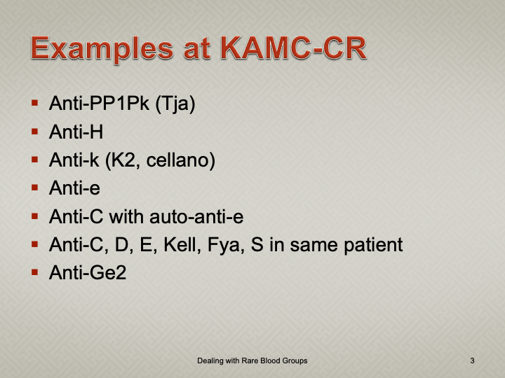
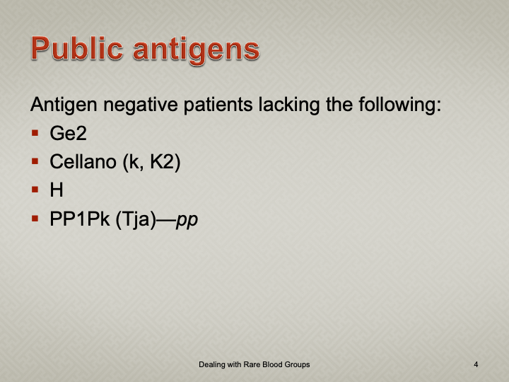
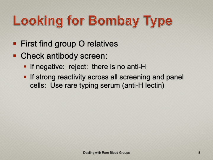
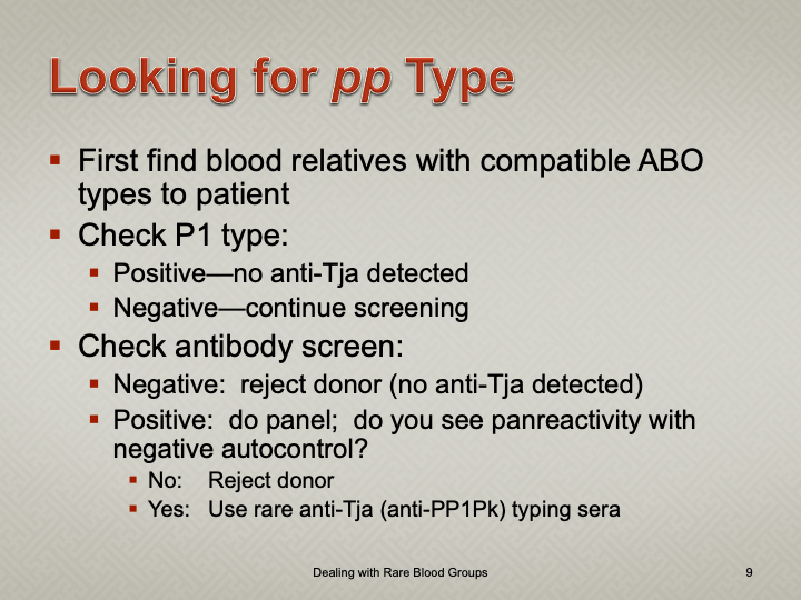
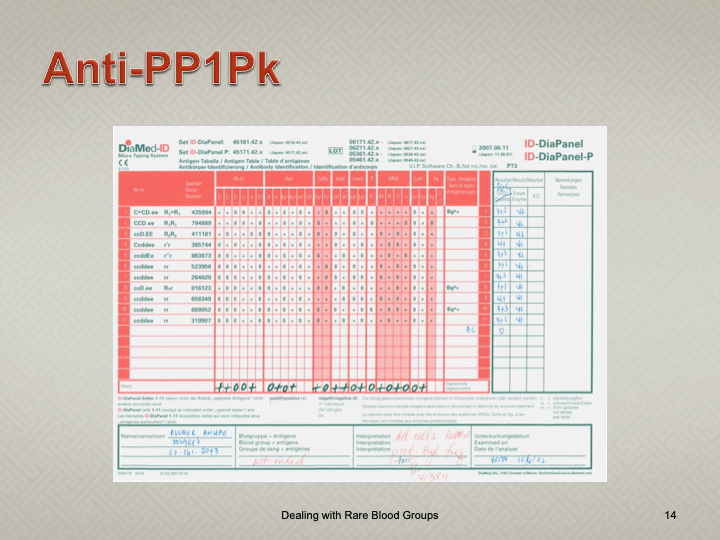
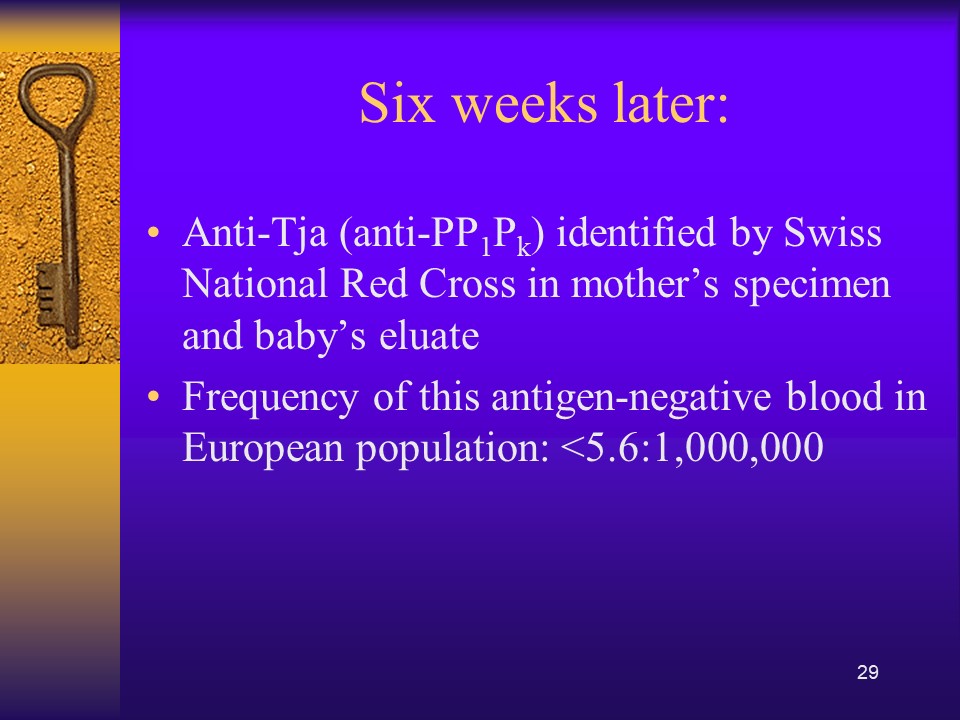
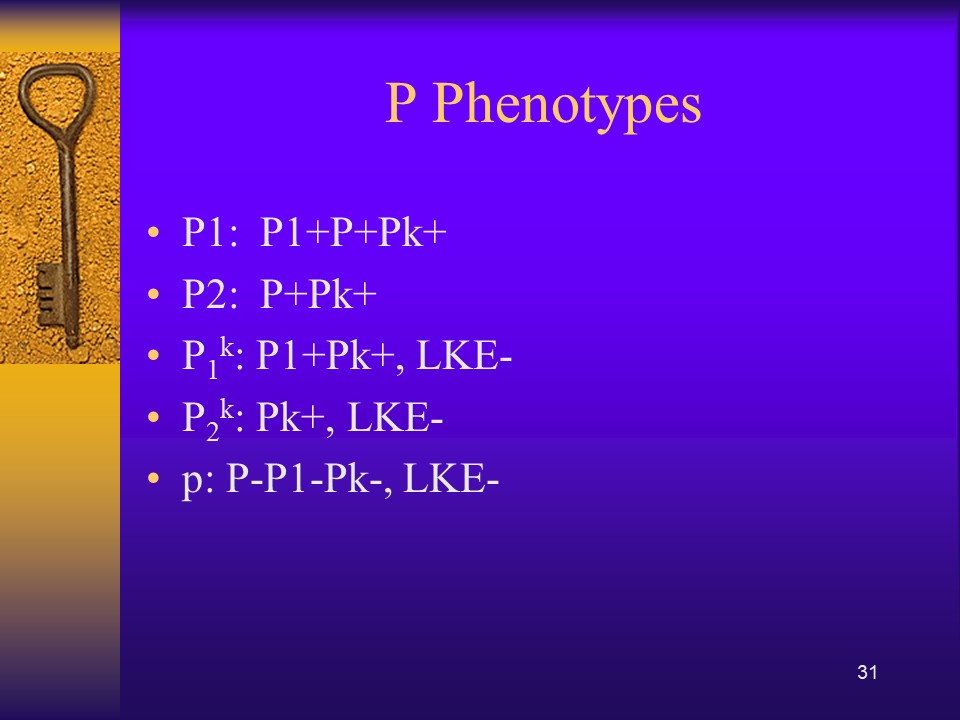
      1. The patient has a dangerous antibody for which antigen-matched units cannot be easily obtained (e.g. anti-k (cellano), anti-PP1Pk (anti-Tja), anti-H (Bombay and Para-Bombay phenotypes).
    2. Autologous collection of plasma may be considered for patients with IgA deficiency with documented specific anti-IgA antibodies.
    3. Other requests will be reviewed by the Head, Transfusion Medicine or designate.
    4. The final decision to proceed with items 2.1 and 2.2 will be made by the Head, Transfusion Medicine or his designate.
  3. Process for Transfusion Medicine Autologous Procedures
    1. The requesting physician shall provide a written or electronic order to the Blood Donor Center.
    2. The request will be reviewed by a transfusion medicine physician.
    3. If rejected, the requesting physician will be notified with the reason for the rejection.
    4. If approved, the donor shall be screened by the usual donation process except:
      1. Hgb >= 11 g/dl will be acceptable for whole blood collection.
      2. Females may also donate autologous plasma.
      3. The last autologous donation will be at least 72 hours before the elective procedure.
    5. Marker testing:  Components from autologous donors with confirmed positive cases of HBV, HCV, or HIV will NOT be used for autologous donation and will be destroyed.
    6. Computer:  Autologous collections will be entered as specifically at the time of registration in the Medinfo  Hematos IIG blood bank computer system and be labeled as autologous in their corresponding ISBT labels.
    7. The transfusion criteria for autologous units shall be the same as for homologous blood.
    8. Both autologous and allogeneic units will follow the same compatibility testing algorithm.
  4. Responsibilities:
    1. Predeposit:  Directly under the control of Transfusion Medicine for all aspects:  policies, procedures, and direct performance of the procedures, including annual review of criteria
    2. Perioperative:  Division Head, Transfusion Medicine involved in conjunction with Surgery and Anesthesia through the National Transfusion Committee.
    3. Intraoperative:  Division Head, Transfusion Medicine involved in conjunction with Surgery and Anesthesia through the National Transfusion Committee.
    4. Postoperative:  Division Head, Transfusion Medicine involved in conjunction with Surgery and Anesthesia through the National Transfusion Committee.

References:

  1. Standards for Blood Banks and Transfusion Services, Current Edition, AABB, Bethesda, MD, USA, 2014
  2. TRM.41600 CAP Checklist Standard

Rules in Medinfo Hematos IIG Blood Bank Software

Medinfo Hematos IIG has an underlying framework of functionality.  It is flexible since it acts upon rules based on parameters (e.g. sex, age, diagnosis, test results, etc.)  You can change the processes in the system by changing the parameters without upsetting the underlying structure of the software.  This means you can make changes very simply and quickly without having to “hard-code.”

Rules are based on parameters which are entered into the system by the user or the results of previous action.  I am listing here some examples of parameters used to define processes in my Medinfo installations:

  • Demographics (e.g. age, sex, nationality, address)
  • Diagnosis
  • Ordering Physician
  • Project (e.g. research project)
  • Location (e.g. hospital ward or clinic)
  • Procedure (e.g. apheresis, surgical)
  • Test results (ABO typing, DAT, marker testing, control values)

Based on these parameters, the system may require:

  • Specific selection of component, derivative
  • Special processing of component
  • Selection of specific test methodology
  • Discard of blood component
  • Invalidation of test rests (e.g. positive D control)

Examples:

  • Restrict use of CCP if SARS-CoV-2 antibody titer is low
  • Discard component if HBsAb quantitation below a threshold (e.g. 100 IU/liter)
  • Hematology ward patients only to receive irradiated blood components
  • Patients in research project to have special testing always done
  • Patients with unexplained hemolysis to have special DAT performed (IgG, IgA, IgM, C3c, C3d)
  • If anti-Kell present, only use K-negative blood—if Kell positive or Kell untested, block allocation
  • If female donor, whole blood only to be processed for RBCs
  • Reflex testing (if HBcAb reactive, then HBsAb must be performed)

Overriding rules:

One can also define if the rules can be overridden by someone with appropriate credentials:

  • Use antigen-incompatible RBCs (e.g. C-positive in a patient with anti-C)
  • Use of non-irradiated RBCs if irradiator is broken

On the other hand, rules can be specified to prohibit overriding:

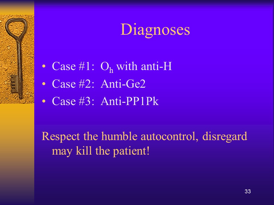
  • Use of group O RBCs in a patient with anti-H

Teaching Document: Variances in Transfusion Medicine

Principle:

In accordance with AABB Standards, all actions contrary to the standard operating procedures and policies of Transfusion Medicine must be specifically approved by the Head, Transfusion Medicine or designate.

Documentation of variances must be organized in a system for ready retrieval for analysis.  They should not be entered into a system that is cumbersome to find the entered variances.

Examples include but are not limited to:  Rh(D)-incompatible transfusions, least-incompatible crossmatch, extension of expired rare reagents, etc.

If the same variance is occurring frequently, it should be determined if modifications in the underlying documentation (policies, processes, procedures) should be made.

Policy:

  1. Whenever there is need for a variance in the policies and procedures in Transfusion Medicine, the Division Head, Transfusion Medicine or designate must be informed.
  2. The Division Head or Designate will review and accept/reject the request.
  3. If accepted, the variance must be documented in writing by any of the following:
    1. A paper form (pre-blood bank computer system implementation) that can ultimately be scanned or an electronic version stored in Transfusion Medicine.
    2. A comment in an appropriate field in the Medinfo computer system
  4. All verbal authorizations for a variance (e.g. telephone call to Transfusion Medicine physician at night) must be recorded on a form and submitted to the responsible TM physician for review and signature.
  5. All variance documentation must be readily retrievable for analysis.
  6. All variances must be collated and assessed as part of the monthly quality review.

References:

  1. Technical Manual, Current Edition, AABB, Bethesda, MD, USA
  2. Standards for Blood Banks and Transfusion Services, Current Edition, AABB, Bethesda, MD, USA

Weak D Phenotype

I prepared this policy for HMC Qatar, which did not perform genotyping.

Principle:

Weakened expression of the D antigen may be due to either quantitative or qualitative defects.  Qualitative defects (partial or mosaic D) are uncommon.

Patients with quantitative defects have the whole D molecule and will not make anti-D upon exposure to D-positive RBCs.

Patients with partial or mosaic D may form antibodies against the portion of the D molecule they lack.  However, not all partial D types may make anti-D upon exposure.  Since we cannot specifically test for this condition, we shall consider females of child-bearing age with weak D as potentially partial D and use D-negative RBCs.

Genotyping for the D gene may define better which partial D types need to receive RhIG or D-negative transfusions.

Definition:

Weak D is defined as weak (1+ or weaker) reactions with one or more routine reagents or reactions only at antiglobulin phase (i.e. Du testing).

Policy:

  1. All donor units must be tested by a method to detect weak D phenotype.  If such testing is positive, the donor unit must be labelled as D-positive and be used routinely for D-positive patients.
  2. Weak-D females of child-bearing ages (<50) should receive D-negative RBCs.
  3. Weak-D males may receive D-positive RBCs unless they are a potential recipient of chronic transfusions (e.g. thalassemia, sickle cell disease, etc.)

References:

Technical Manual, Current Edition, American Association of Blood Banks, Bethesda, Maryland, USA

Standards for Blood Banks and Transfusion Services, Current Edition, AABB, Bethesda, MD, USA

Issues with Labels

You can have the most sophisticated blood bank software, but if you can’t read the labels or if they fall off, you have a disaster.  These are my thoughts from implementing our blood bank computer system back in 2013.

Check Digits:

Both ISBT specimen and product labels have an internal system to verify that they have been read correctly.  Within the blood bank software, this should not be a problem.  However, can your third party such as a hospital information system HIS read them?
ISBT Compatibility:

The institution’s HIS could not read the component labels.  To this date, the problem has not been fixed.  As a workaround, we sent them the ISBT label codes directly from the blood bank software.  The only complete transfusion record was in the dedicated blood bank computer system, not the HIS.  You could not rely on the bedside nursing entry at all.

The HIS did not use the ISBT database and had no values for the E codes.  Again, we had no choice but to send an abbreviated E-code descriptor to them.  We did not use their transfusion module at all, but one of our clients did.  They had to hard code the list of E codes in use with their descriptors into their HIS.

Label Adhesive

We tested candidate labels at room temperature, 1-6, minus 18, and minus 80 C.  We found that most of the labels’ adhesive were not sticking at minus 80.  For some, you could literally blow on the blood bag and the label fell off.  I imagined a scenario in which I opened a freezer and saw the blood bag labels all lying separately at the bottom.

RFID Tags:

Do you use an RFID tag integrally attached to the ISBT label OR do you stick a separate RFID tag?  If the latter, how do you ensure that you put the proper tag on the proper bag?

Readability:

Readability:  Can all your blood bank devices read your printed labels?  Do you have to adjust the printers for this?  Whose responsibility is it to do this? 

Labels printed outside the blood bank:

If you receive patient specimens from outside the blood bank, can your devices read them?  Who is responsible to adjust the printers in the wards and clinics?

Validations:

Who validated that the HIS prints the accurate complete label for the right patient?  We discovered that this was not the case with our HIS and needed correction by them.  Remember that any processes affecting Transfusion Medicine should be assessed by Transfusion Medicine.  Do not accept verbal assurances from anyone, not even your HIS.

2/2/21

Teaching Document: Validation Process

This is a teaching document for medical technology and transfusion fellows to explain the general structure of a validation.

Principle:

All validations must be planned.  A validation protocol must be prepared with specific criteria for acceptance.  All validations with attached evidence must approved by the Head, Transfusion Medicine.

Policy:

  1. A written validation protocol must be prepared in the advance and at least including the following:
    1. Specific parameters and number of iterations to be performed
    1. Designated staff to perform validation
    1. Documentary evidence of the testing
    1. Specific acceptability criteria
  2. The completed validation protocol must be submitted to the Division Head, Transfusion Medicine, or designee for review.
  3. Once the validation plan has been reviewed, it must be performed by the designated staff.
    1. Software validations will be performed in a specific test environment, not in the live, production system.
  4. The completed validation document, including screenshots of the software functionality if applicable, must be submitted to the Division Head, Transfusion Medicine for review.
  5. The equipment or software may only be used if the acceptability are met AND the validation is approved by the Division Head, Transfusion Medicine or designee.
  6. The completed validation protocol will be stored in the document control system.

Reference:

Standards for Blood Banks and Transfusion Services, Current Edition, Bethesda, MD, USA

Blood Component Variances

Principle:

AABB Standards requires that all variances are documented and investigated and corrective actions taken when necessary.  Any time a blood component is found to be defective (e.g. broken seal, leaking, discoloration, clots, etc.), mislabeled, or testing results incomplete or not documented,  the cause should be investigated by the Donor Center and reported back to the initiator of the report in writing.
Policy:

  1. All transfusion services must inspect all blood components upon receipt (e.g. for leakage, broken seals, improper temperature, clots, discoloration, gas, etc.).
  2. Labels must be compared to the consignment sheet for complete concordance.
  3. If units are found that are not listed or mislabeled, they must be reported in writing to the Donor Center and returned as-is for investigations.
    1. If the unit is leaking or broken, ensure standard/universal precautions are taking to minimize contact with the fluids.
    2. Damaged blood components must not be used.  Units with mislabelings or other discrepancies between the labels and the consignment sheets may be used when such errors are corrected and officially reported by the Donor Center.
  4. Use the standard incident (occurrence variance) report form (OVA) for each and every variance.
  5. The submitting location should keep a copy of the OVA and immediately forward the original to the Transfusion Quality Section.
  6. The Donor Center should investigate the variance and prepare a written investigative report and submit to the Division Head, Transfusion Medicine.
    1. Donor Center investigations should be completed within one calendar week.
  7. The Donor Center should forward a copy of the completed written investigation to the transfusion service which initiated the investigation.
  8. The copy of the investigation report should be attached to the OVA and kept at the local site.
  9. Transfusion Quality shall include these variances in its monthly reports.

References:

Standards for Blood Banks and Transfusion Services, Current Edition, AABB, Bethesda, Maryland, USA

Washed RBCs

Note:  This is an updated version of a previous post.

Principle:

Washing RBCs removes plasma and reduces the leukocyte count only by 1 log.  For leukodepletion, we must rely on filtration to reduce the WBCs to less than 1 x 10⁶ per unit according to CE rules.  Red cells or platelets in additive solution contain only minimal plasma (about 35 ml).  There are few definite indications for washing RBCs and it should be rarely necessary.

Policy:

Washing RBCs should only be done in the following circumstances:

  1. Deglycerolization of frozen RBCs.
  2. Severe allergic or anaphylactic reactions to plasma proteins
  3. IgA deficiency with anti-IgA
  4. Paroxysmal nocturnal hemoglobinuria PNH—relative indication (often these patients receive RBCs before the diagnosis of PNH is confirmed)
  5. Transfusing a previously irradiated RBC unit for pediatric use if more than 24 hours has passed since it was irradiated.
  6. Any other time when so designated by a transfusion medicine consultant.

Note:

  1. If anyone requests washed RBCs and it does not fit into one of the above categories, contact the transfusion medicine consultant.
  2. Washed RBCs are NO substitute for leukodepleting RBCs by filtration NOR can they be used in place of irradiation for prophylaxis against transfusion-associated-graft-versus-host disease TAGVHD.  Using the Reveos automated component processing system, all components are leukodepleted—RBCs are released in SAGM.

Reference:

Standards for Blood Banks and Transfusion Services, Current Edition, AABB, Bethesda, MD, USA

Opinion: Ready after Fellowship?

I was recently interviewing a candidate for consultant in Transfusion Medicine.  Several months previously he had completed a fellowship in Transfusion Medicine in the United States.  He was applying for a position in my hospital in Qatar, which included seven hospitals and a blood donor center.  He had no training in donor management or therapeutic apheresis.

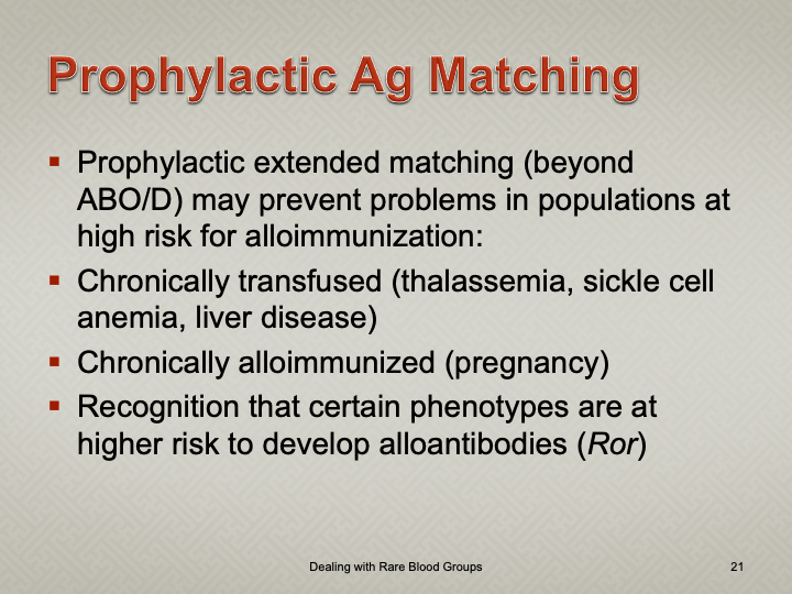
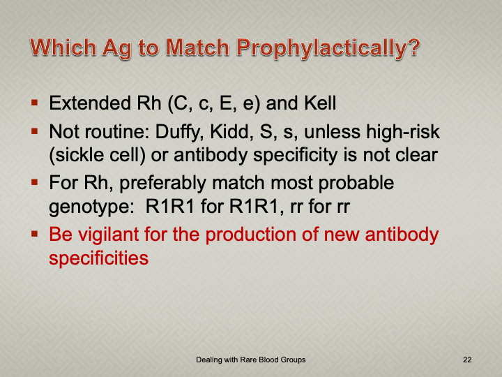
The successful candidate was to rotate on-call to cover all hospitals and the blood donor center.  He had never worked outside the United States.  Routinely, he did not review antibody panels since those workups were usually sent to the local blood provider there.  In his training, he had strictly followed US FDA and American version of AABB Standards.  His training center did not routinely do extended phenotypes (C, c, E, e, and Kell).  Extra testing and phenotyping had to be explicitly ordered by the clinician to get reimbursement.  Thus, there was no prophylactic antigen matching done on patients.  He did not feel comfortable reviewing antibody panels.

He had no experience with universal leukodepletion, pathogen-inactivation, platelet additive solutions, or automated component production such as the Terumo BCT Reveos.  He did not interpret donor marker testing results.

On the contrary in our organization, the transfusion medicine physician had to review all antibody panels (usually he was the most knowledgeable person for this).  We followed the Council of Europe CE and other practices that did prophylactic antigen matching.  We were also in charge of donor qualification and therapeutic apheresis and reviewed any product deviations from the Reveos and donor marker testing.

Clearly, this candidate did not practice transfusion medicine in the way that was necessary for our operations.  We could not cut him loose and make him responsible for a hospital transfusion service or the blood donor center.

Let us contrast this candidate for one being recruited for anatomic pathology/histopathology.  Grossing specimens, performing frozen sections, reading slides, diagnosing cases are the same everywhere in the world.  After completing his American certification, he could perform his profession almost anywhere in the world.

Transfusion medicine practices need to be localized and the selection of blood components and donor qualification are different.  Most of the world does not follow US FDA and has access to blood components, tests, and other technology that is different and maybe more advanced than his training in the USA.

I gave him a clinical scenario to interpret.  An AB patient with anti-K needs to be transfused with plasma.  Are there any special requirements for the plasma?  What if the only AB donor had anti-K would you use it?  What if the only RBCs available had not been phenotyped for Kell?  What would you do?

He did not know that we discard plasma with clinically significant alloantibodies routinely.  He did not want to phenotype the RBC unit for this patient since this had not been explicitly ordered by the clinician.

My recommendation was not to hire this candidate if there were others who had worked in European or similar systems to our own practices.  In effect, to use this physician, he would have to undergo a mini-fellowship to learn our practices since they were contrary to ours.  Unfortunately, we were very short-staffed and did not have resources to offer this training.

In summary, blood bank practices are very localized.  If you are considering to hire staff from other countries not following your standards, you must assess if the candidate is flexible to change his practices and/or whether you have the resources to train the physician.