Antibody Titration including Software Processes

Processes and Software Building—Part 20

My practice across the globe has exposed me different rationales to performing antibody titration.  In my American training and practice (and also at international institutions following the American version of AABB accreditation), I only routinely performed titration of anti-D for Rh(D) hemolytic disease of the newborn and anti-A/anti-B for ABO-incompatible stem cell transplants AND ABO-incompatible renal transplants.

I have had heated arguments with some physicians who insisted they wanted titers for other antibodies.  The AABB Standards do not require this but leave it to the discretion of the Transfusion Service Medical Director.

In my entire career, I never worked in a blood bank or blood center which had optimal staffing or resources.  I focused on what was medically/technically necessary and even then still had shortages.  If performing a test will not change the clinical treatment, why perform it unless you are doing a research project!

Titration is a time-consuming, and until recently, a tedious manual task.  Recently some of the automated immunohematology analyzers offer a titration program.  We used the Ortho Vision Max which could perform both IgG and IgM titers within one hour—walk away!!  However, during that time, the titration procedure monopolized the analyzer.

The ABO-incompatible renal transplant program at HMC Qatar was modelled after Sweden’s Karolinska Institute.  However the latter site performed manual IgG and IgM titrations using Biorad/Diamed gels.

I did not have sufficient resources to commit staff to manual titration at HMC so I did a comparison study between the Ortho Max and the Biorad methods.  We were able to get good correlation and used the automated method for the transplant.

I am still biased against performing titrations for other antibodies.  I always ask, ‘Does the titration correlate with clinical severity?’  Unlike anti-D, antibodies such as anti-Kell and anti-c may be low titer but cause death.  Can anyone show me a definitive study that titers are useful except for transplants and Rh(D) hemolytic disease of the fetus/newborn?

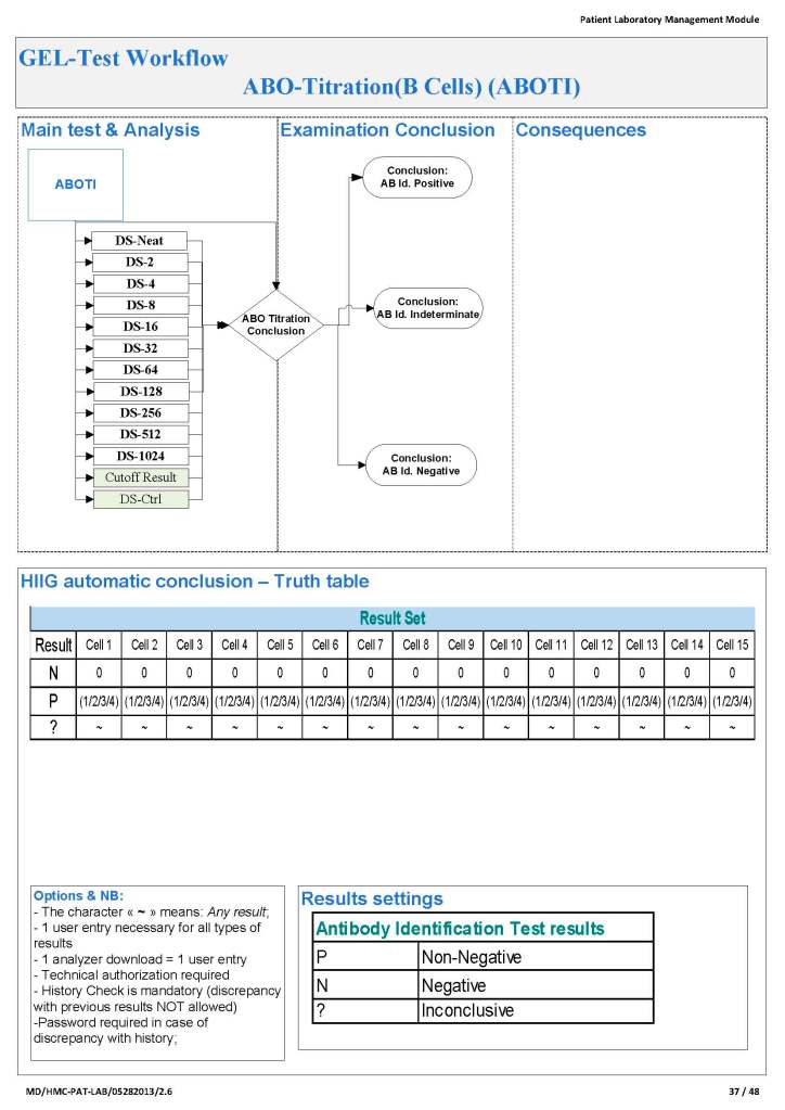
Since the method was working well on the Ortho equipment, I next established an interface to Medinfo.  The test was performed separately for IgG and IgM antibodies.  Medinfo recorded the reactions in all the wells.  The last well showing a 1+ reaction was interpreted as the titer (e.g. if 1:64 were the last 1+ reaction, then the titer was 64 in Medinfo).

The Medinfo process is shown below.

20/7/20

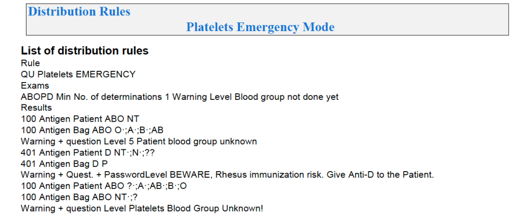
Platelet and Plasma Allocation Rules

Processes and Software Building—Part 20

Medinfo software is rules-based so the institution may set its own custom rules for all processes.  One chooses a framework and then adds any additional rules it needs for optimization.  Turnkey systems do not offer this flexibility.

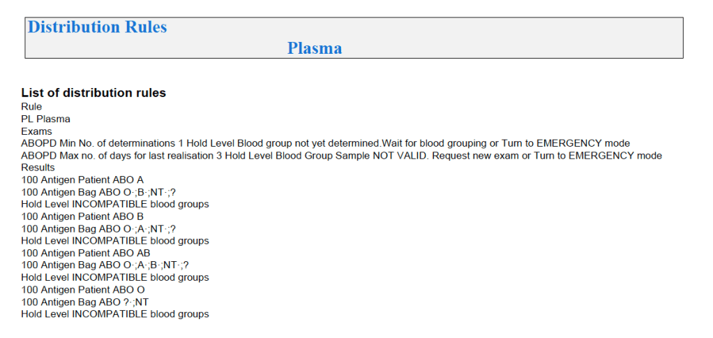
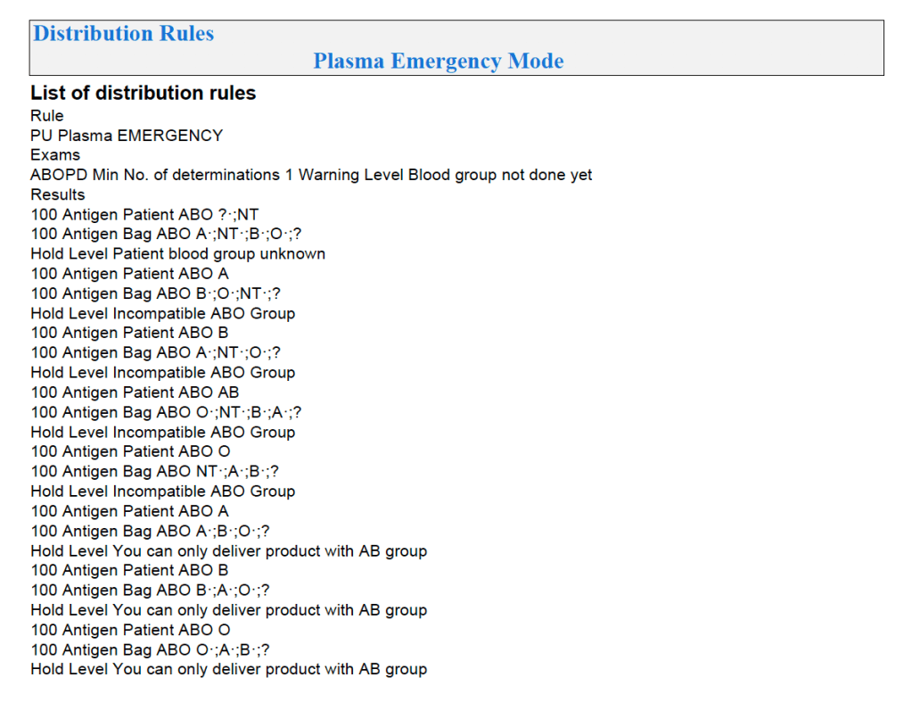
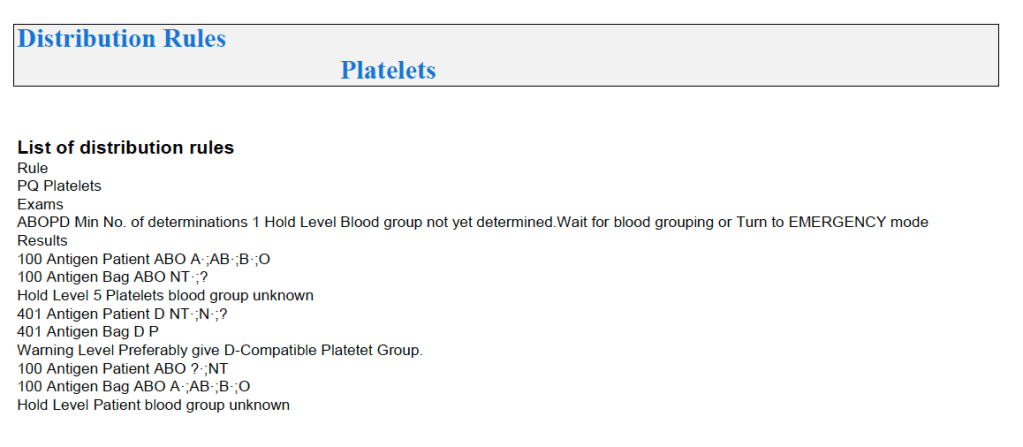
The rules for platelet and plasma components are much simpler than those for RBCs since usually we only consider ABO type.  There are two modes:  regular and emergency, the latter applying if not all the patient testing (including historical checking) is available.  The components, on the other hand, must meet all criteria before being considered for patient use.

Example rules for plasma follow:

For platelets, note that for adults and anyone else >= 20 kg, I gave any type of platelet pool or plateletpheresis component without regard to ABO matching.  With our production method, I did not give Rh immunoprophylaxis to females of child-bearing age receiving platelets from D-positive donors based on our clean (essentially RBC-free) Reveos automated production process.

Similarly, allocation rules for granulocytes, etc. could be made and enforced by the software.  Low-B-titer group A universal plasma would also be easy to implement.

To Be Continued:

19/7/20

RBC Antigen Matching and Software Processes

Processes and Software Building—Part 19

In the previous post I outlined how Medinfo handled antibody screening and identification.  This post reviews how antigen matching is used based on these results.

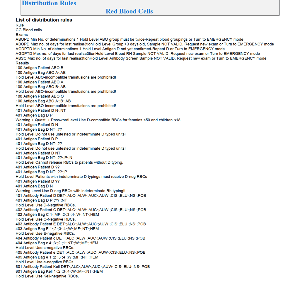
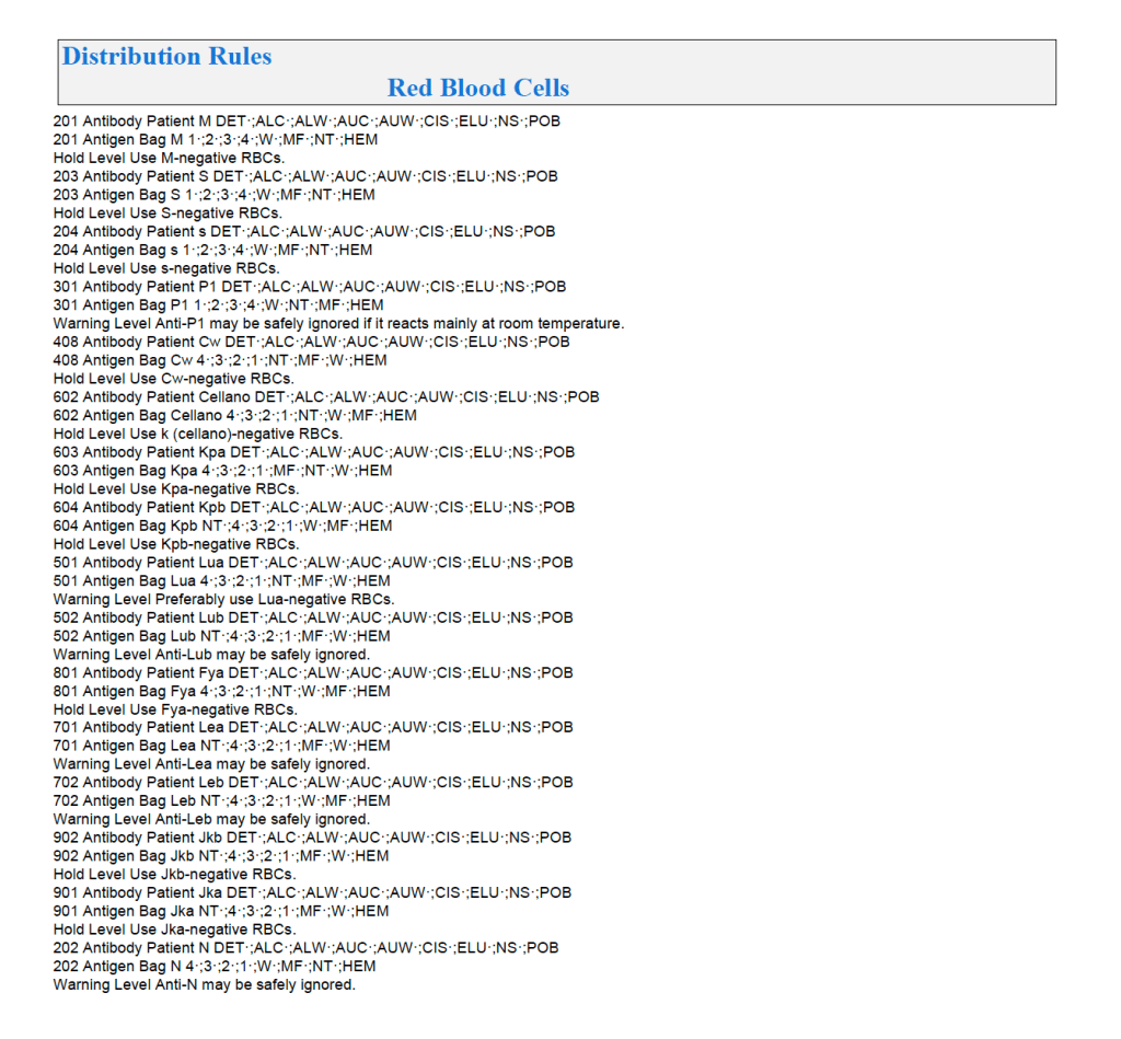
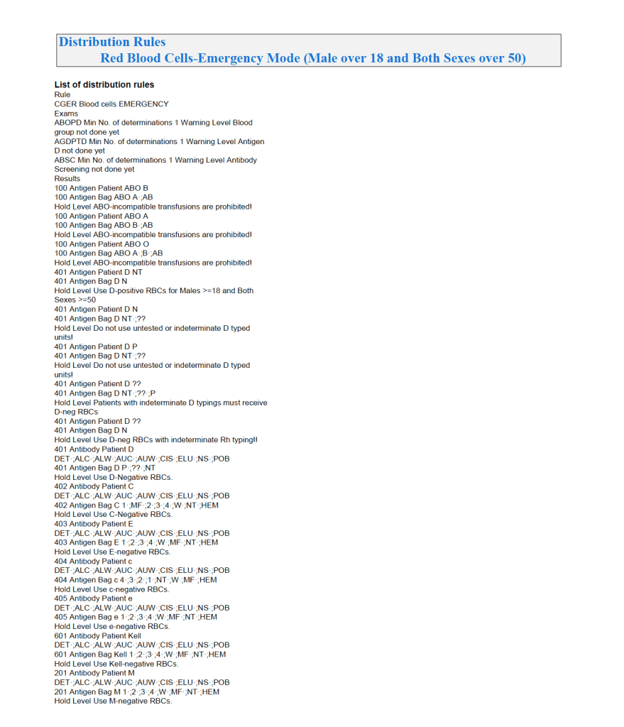
There are two modes, regular and emergency.  If the patient has not had at least two ABO/D determinations and/or does not have a recent antibody screen, then emergency mode must be selected with its own rules.  Otherwise, the regular mode applies.

Regular Mode:

In general, if there is a clinically significant antibody, an RBC unit which has not been matched for the corresponding antigen or has the corresponding antigen cannot be routinely selected.  However there is a hierarchy here also:

  1. Absolutely prohibited release—no one can override the logic (e.g. giving group O to a patient with anti-H)—not even the transfusion medicine physician can override this
  2. Restricted release—only certain staff can release the incompatible or untested unit (e.g. giving C-positive unit to someone with anti-C)
  3. Least-incompatible for WAIHA:  requires transfusion medicine physician approval
  4. Informational release:  authorized staff may release antigen-incompatible or untested unit but a pop-up menu appears and asks them to accept (e.g. Lewis untested unit in a patient with anti-Lea).
  5. Antigen-specificity matched—the usual mode for patients with antibodies

Examples of Regular Mode rules follow (these are not the complete lists but just provided to show the complexity of the process).

Emergency Mode:

This is much more restricted for selection of ABO/D and other antigen typings.  An example follows:

A similar hierarchy exits for platelet and plasma allocation and will be considered in a future post.

To Be Continued:

18/7/20

Antibody Identification and Software Processes

Processes and Software Building—Part 18

The blood bank is one of the last bastions of manual testing, especially for complex procedures.  Basic antigen typing, direct antiglobulin tests, and antibody screening adapt well for routine automation, but the actual antibody identification requires multiple methods and requires considerable experience to complete. At least today, a machine cannot replace the blood banker!!

One could take the approach to perform antibody panels in an automated system and record the results.  However, these are worthless unless you have the actual panel in hand.  Artificial intelligence to analyze the panel results is limited.  You still have to manually review everything.  In Medinfo, we could also scan the manually performed panels and add them to the record.

While I was at HMC, some of the hospital transfusion services elected to run panels automatically and then review the results manually.  We prepared a contingency to record the panel results but these by themselves could only be used with the actual red cell panels in use.

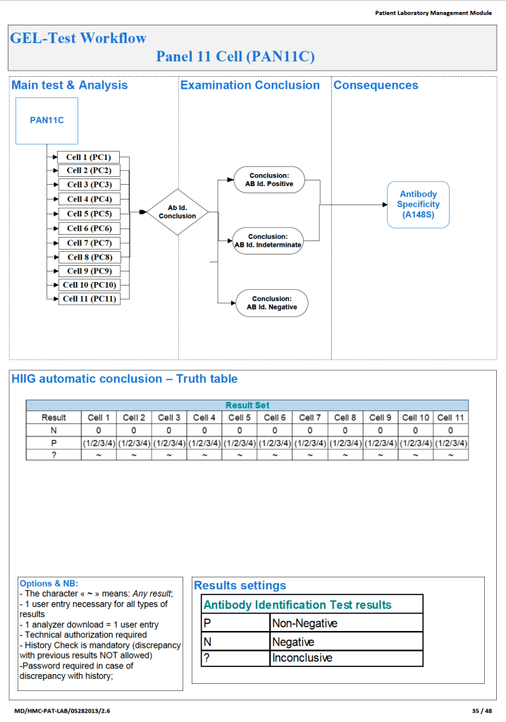
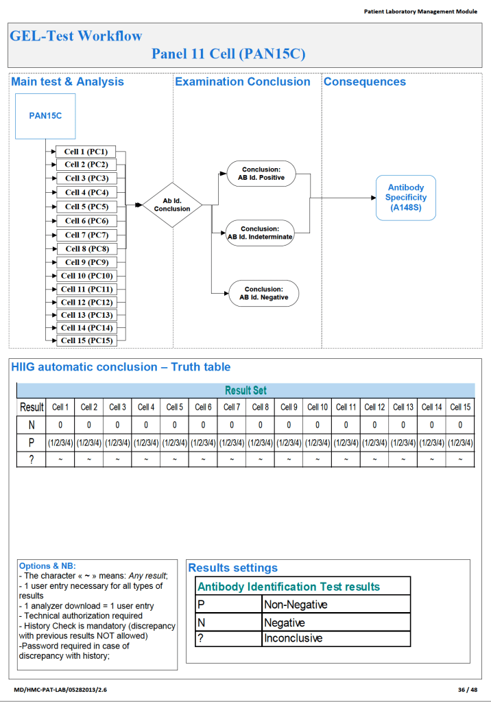
Examples of 11-R and 15-cell panel result importation follow:

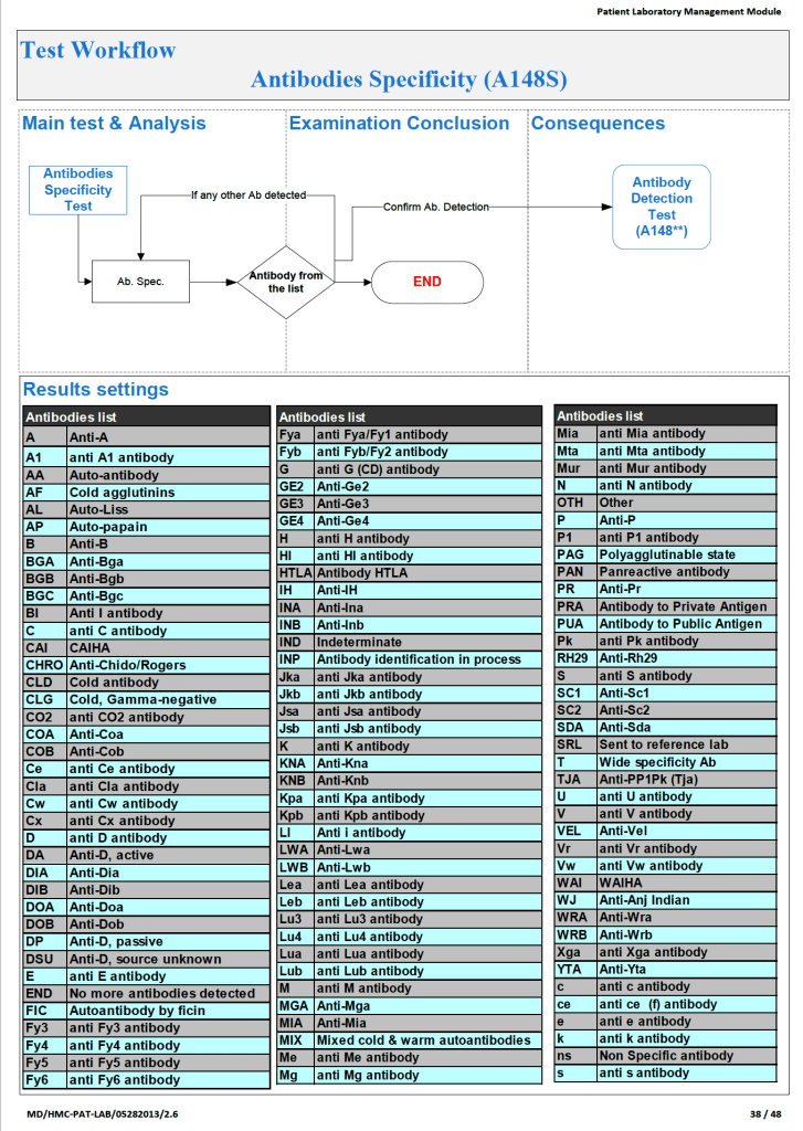
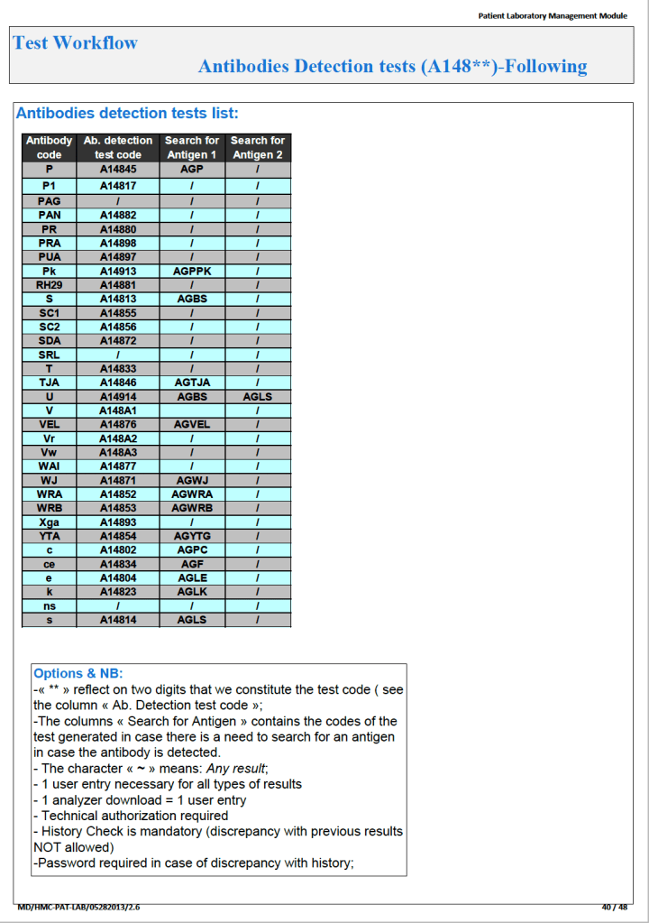
What we absolutely had to do was enter the antibody specificity in a way that it could trigger our algorithms, i.e. our rules-based system of RBC allocation according the permissible blood types.  Each antibody specificity and each antigen typing had its own unique code.  It could not be entered free text!  Workflows follow:

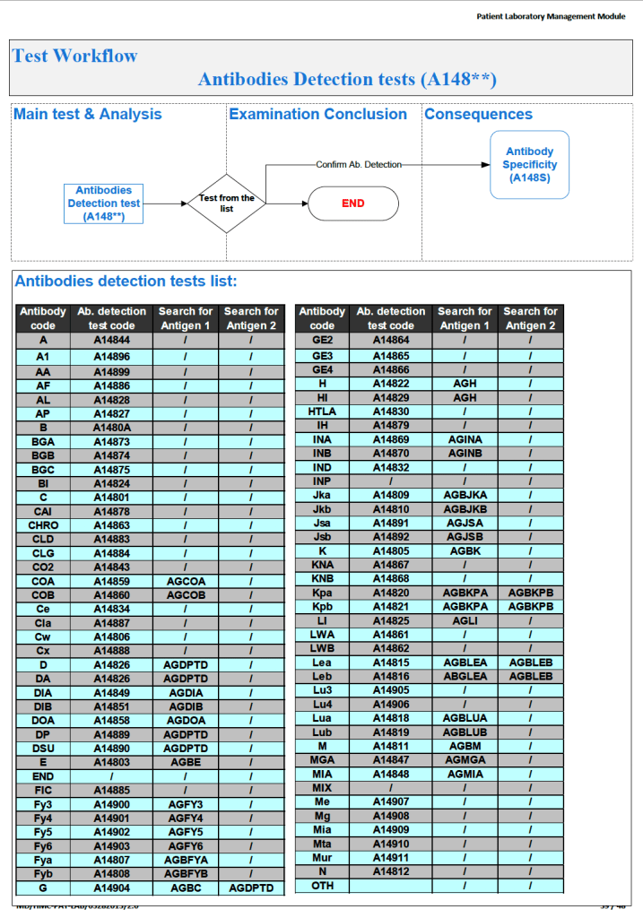
Next, the antibody specificity test would trigger antigen typings for the correspond antigen and/or closely related antigens:

Based on the antibody specificity and antigen typings, a whole series of rules for routine and emergency-mode release were triggered.  This will be discussed in a subsequent post.

To Be Continued:

17/7/20

Elution Indications and Software Implementation

Processes and Software Building—Part 17

The direct antiglobulin test DAT may be the first or last place that a delayed hemolytic transfusion reaction can be detected.  In my practice, I always performed it the first time I encountered a patient with a positive DAT.  If the subsequent DAT testing is of the same strength and there is no change in the clinical status of the patient, then I might empirically repeat the elution after 14 days, i.e. just when new antibody emergence may be detected.

In summary, my criteria for elution after a positive DAT are:

  1. First patient encounter with a positive DAT
  2. At least 14 days since the previous elution
  3. DAT strength has increased since the previous exam (at least 1+ increase in strength in either IgG or C3)
  4. Patient shows evidence of hemolysis
  5. Suspected cases of drug-related hemolysis
  6. Transfusion Medicine physician specifically orders it otherwise

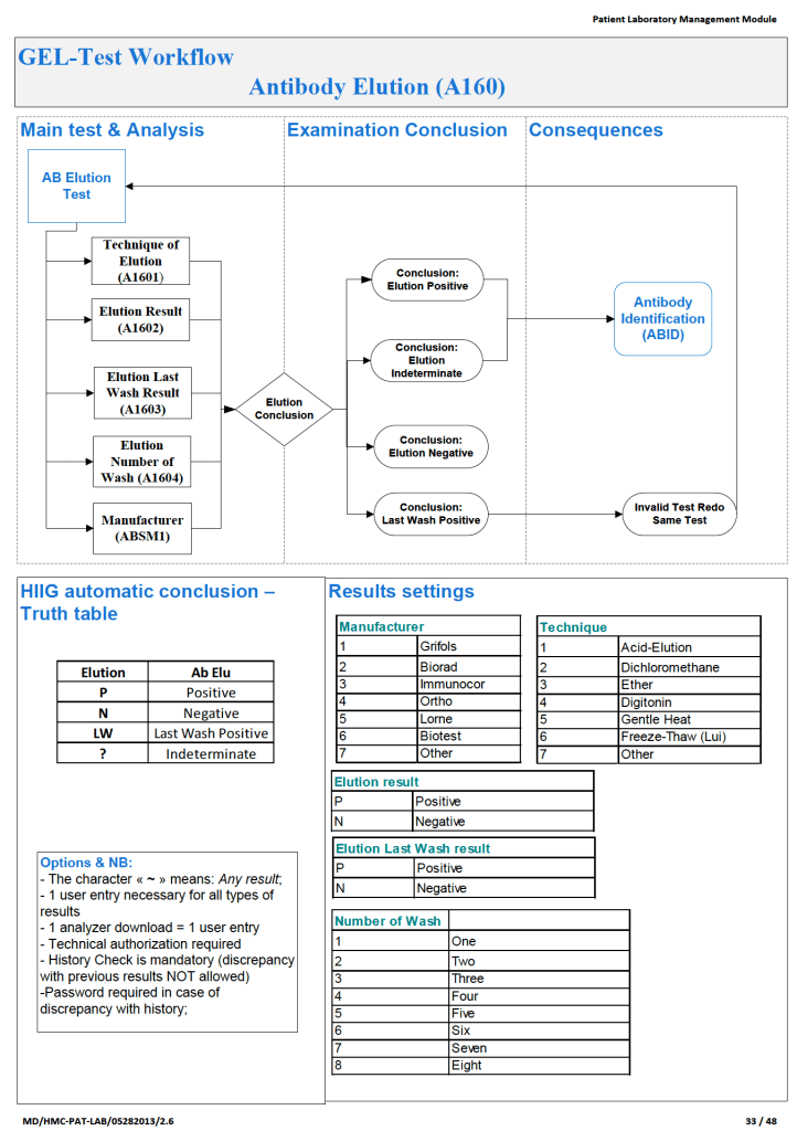
In the software processes, the following parameters were recorded:

  1. Technique of elution (acid glycine, dichloromethane, digitonin, ether, etc.)
  2. Number of washes (too many may remove the antibody from the RBCs)
  3. Last wash result (must be negative to accept conclusion)
  4. Manufacturer of the elution kit (if any)
  5. Elution result based on panel testing

Note:

  1. All indeterminate or positive eluates would trigger an antibody identification.
  2. All results would be reviewed by me or the covering Transfusion Medicine physician.  A comment would be entered into the results that could be viewed by the clinician either directly from Medinfo or through the hospital information system.

If the DAT was only positive for complement, it was up to the Transfusion Medicine Consultant to decide whether to proceed with elution.  Personally, I would usually proceed because the eluate is more concentrated and may occasionally detect an antibody that was not noted in the patient’s plasma.

The attached Medinfo workflow shows the process designed by me for use at HMC Doha.

Processes and Software Building: Updated Convalescent COVID-19 Plasma Production

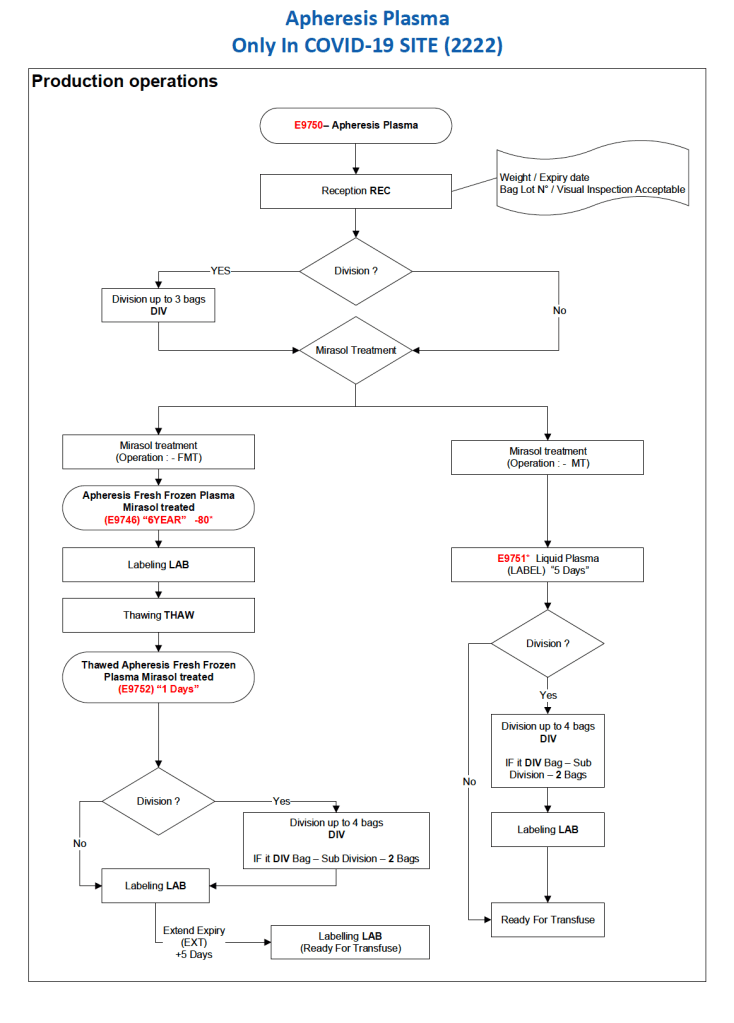
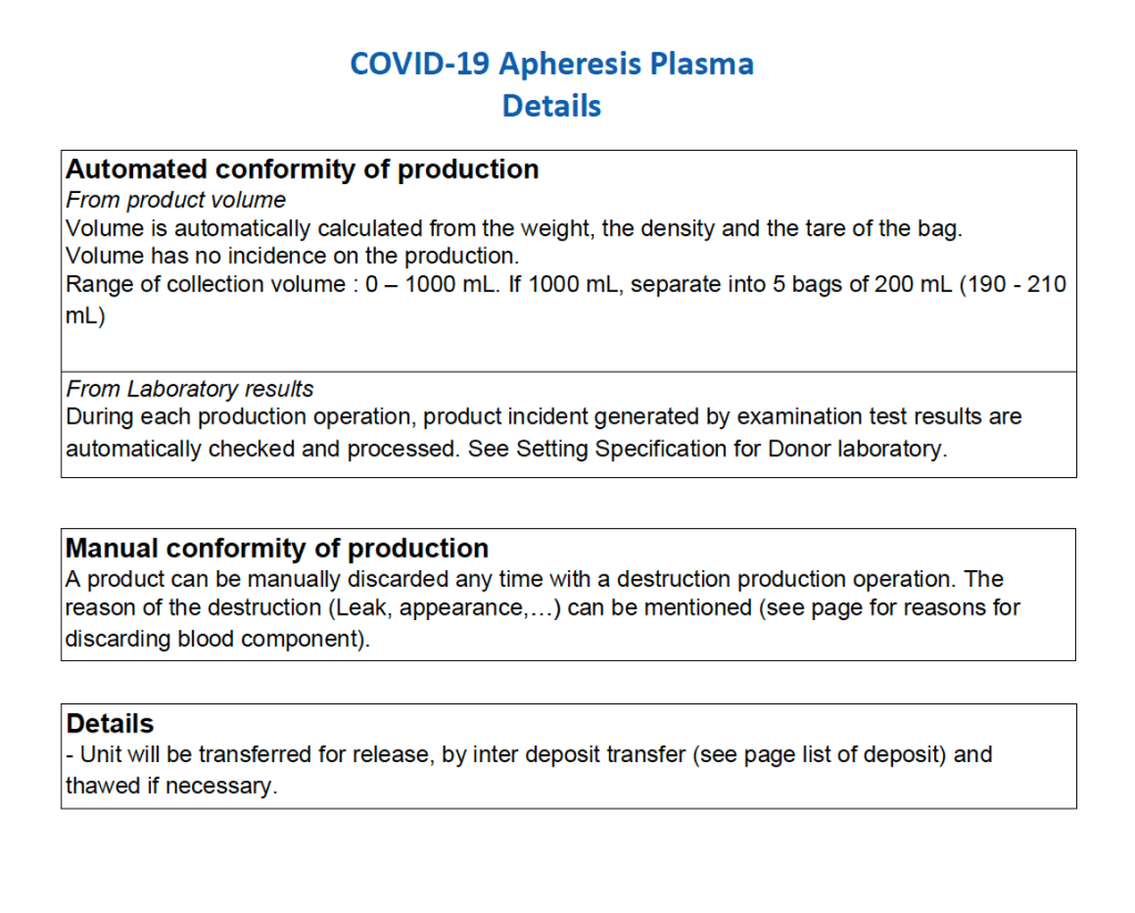
After the initial manual setup of the CCP program, the Medinfo process was set up.  The following workflow shows the production of CCP from the raw apheresis collection, including division into aliquots based on the total volume.  The plasma volumes were kept within the range for riboflavin pathogen inactivation (Mirasol).

The usual safeguards for production were also in effect for CCP.  The product could not be labelled without all criteria (donor screening, collection, marker testing) being met.  Furthermore, the inter-depot and transfusion service processes still applied.  However, all steps were done in quarantine at a location separate from the regular processes.  Also, the actual ordering and release of CCP was restricted to the quarantine hospital blood bank site.

The following outline the production process:

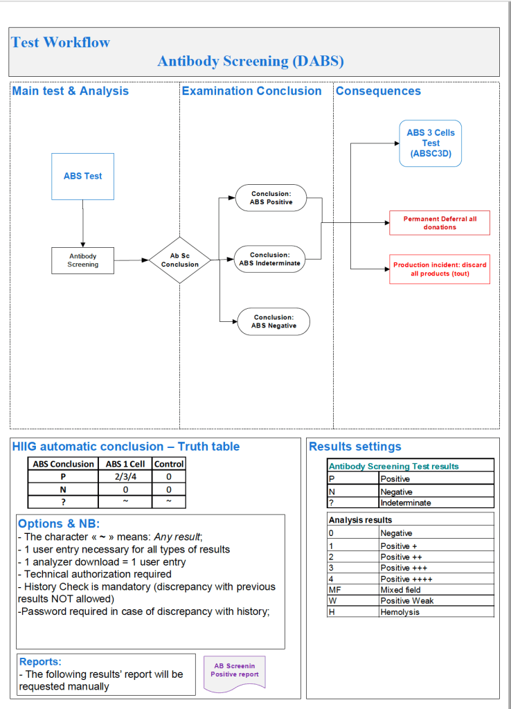
Software Processes for the Antibody Screen

Based on the previous software post, I use the antiglobulin test algorithms to construct the antibody screen ABS, namely an INDIRECT antiglobulin test, i.e. incubate reagent red cells with the donor or patient’s plasma and then perform the DAT.  At HMC Doha and at NGHA in Riyadh, we mainly used three cell screens (no pooled cells for donors). The reactions could be machine-read (we had the interface) but the interpretation of the specificities found was done manually.

Donor ABS:

We screened all donors for irregular antibodies.  I disqualified most donor with non-negative screens from donation (my personal preference).  However, for nonspecific antibodies, I might elect to discard the current collection and then reassess the donor’s status at the next encounter.  Not everyone would agree with this most restrictive approach but I would not use the platelets or plasma in such situations.  I also did not use the donor RBCs, even though they were in SAGM—but I could elect to override this decision in Medinfo if there were a rare blood group (e.g. r’r’).

Example #1 (following) shows a sample donor ABS process:

Patient ABS:

Excluding emergency release, if the patient did not qualify for the electronic (computer) crossmatch, then the AHG crossmatch was required.  A three-cell antibody screen including R1R1, R2R2, and rr RBCs was used that usually included cells homozygous for the Kell, Duffy, Kidd, MNS antigens.  For any case with a non-negative antibody screen, either donor or patient, an antibody identification was performed.

Note that I did not use any other software to make antibody identifications.  I required my technologists to be proficient in identifying basic and intermediate-level testing.  The actual antigen makeup of each lot number of antibody screen or identification panel was NOT stored in Medinfo.  We scanned the antibody screen and panel workups and could store those in Medinfo.

Example #2 (following) shows a sample patient ABS process:

Patient ABS Fix for Interface Regression Caused by HIS:

Regretfully, with a software update from the hospital information system HIS vendor, a major regression occurred.  It would not discard results from Medinfo if more than one antibody result (e.g. anti-E and anti-c) was sent back.  We could see the results in Medinfo, but those outside Transfusion Medicine could not see antibody results.  The entire interface for antibody identification had to be rewritten so that the HIS would see each result uniquely and thus all results could be sent back to it.

Regression Example:

Anti-Kell sent from Medinfo:  one antibody result, HIS would store and show this result.

Anti-E and anti-c sent from Medinfo:  No antibody results shown, both results kicked out.

Regression Fix:

Anti-E sent as Method Type1-Antibody 1 and anti-c sent as Method Type 1-Antibody 2:  both results displayed in HIS.

Example #3 (following) shows the regression fix.

Software Processes–Direct Antiglobulin Test

Tests and Reagents:

There are many variations for this most important test.  In the very least, this consists of:

  1. Screening polyspecific AHG for detecting IgG and/or complement
  2. If positive, reflex monospecific tests to distinguish IgG from C3 reactivity

There are many kinds of reagents:

  1. Immunoglobulin:  whole molecule IgG, monospecific gamma heavy chain, monospecific alpha heavy chain, monospecific mu heavy chain
  2. Complement:  C3d, C3c, C3b, C5, etc

Regretfully, some of the monospecific are not really monospecific (e.g. whole molecule IgG detects both heavy and light chains and thus may detect IgG and give weaker results for IgM or even IgA.)

Read More »

DAT and Selection of RBC Units for Transfusion

In 1984 effective with the 13th Edition AABB Standards, the requirements for performing a direct antiglobulin test and autocontrol for compatibility testing were eliminated.  The DAT is very important to detect delayed hemolytic transfusion reactions, certain autoimmune conditions, and drug-related hemolysis.

Since that time, the immediate-spin crossmatch and now the electronic computer paperless crossmatch may be used for most compatibility testing in place of the classic, antiglobulin-phase (indirect antiglobulin test) crossmatch.

If an antiglobulin phase (indirect antiglobulin test–IAT) crossmatch is performed, a donor unit with a positive DAT will cause a false-positive reaction.  Since most crossmatching does not include the IAT, it will not be affected by the DAT status of a donor unit.

Policy:

  1. Donor RBC units will NOT be routinely tested for DAT as part of component processing.
  2. The type of compatibility testing selected for a particular patient should be the technically simplest one (no need to do extra work unless so instructed by the transfusion medicine consultant/designate):
  3. Do a full antiglobulin-phase IAT crossmatch if ANY of the following applies:
    1. There are no two independent ABO/D typings on the patient during the current admission.
    2. The ABO/D type of the current admission does not match the historical information.
    3. The patient has a detectable antibody at 37C
    4. The patient has a history of a clinically significant antibody but no current antibody
    5. Whenever the consultant, transfusion medicine/designate requests it.
    6. Whenever the Medinfo HIIG record so indicates (in comment section)
  4. Do the immediate-spin crossmatch if ALL of the following apply:
    1. Only one determination of the ABO/D type
    2. The historical ABO/D type agrees with the current type.
    3. There are no antibodies reacting at 37C AND there is no history of antibodies at 37C.
  5. Use the computer/electronic crossmatch if ALL of the following apply:
    1. There are two determinations of the ABO/D type and they both agree with each other.
    2. The historical ABO/D type agrees with the current type.
    3. There are no antibodies reacting at 37C AND there is no history of antibodies at 37C.
  6. When to do a DAT on a donor unit:
    1. Patient antibody screen is negative but the full AHG crossmatch is incompatible.
    2. Part of a transfusion reaction workup where the AHG crossmatch of donor cells and patient serum is incompatible.
    3. Whenever the consultant, transfusion medicine/designate requests it.
  7. If a donor unit is found with a positive DAT, perform:
    • Do polyspecific and monospecific (IgG and C3) antisera
    • Perform an acid-elution.
    • Send the results to the transfusion medicine consultant/designate for review.
    • The reviewer will enter his review in the Medinfo HIIG in the Donor Consultation Section both as global donor comment and a result-specific comment against the antibody screen result.
    • Use of the DAT-positive donor unit:
      1. Select another RBC unit for the transfusion.
      1. The final decision to use the unit will be made by the Transfusion Medicine consultant/designate.

Important:  Don’t do a classic AHG/IAT phase crossmatch unless you have to do it  (see conditions above.)  A donor unit with a DAT is unlikely to be clinically significant and may be transfused safely to the patient in most situations.  Patients receiving electronic-crossmatch and immediate-spin crossmatch are receiving units with positive DAT without incident.

References:

  1. Standards for Blood Banks and Transfusion Services, Current Edition, AABB, Bethesda, MD, USA
  2. Guidelines to the Preparation, Use, and Quality Assurance of Blood Components, European Committee (Partial Agreement) on Blood Transfusion (CD-P-TS), Current Edition
  3. Technical Manual, Current Edition, AABB, Bethesda, MD, USA

Autologous Transfusion and Responsibilities

This is the process I developed for HMC Doha. The Medical Director (here Head, Transfusion Medicine HTM) is actively involved in the development of policies, processes, and procedures for ALL types of autologous donation in conjunction with the National Transfusion Committee NTC.

  1. Predeposit:  Directly under the control of the HTM for all aspects:  policies, procedures, and direct performance of the procedures, including annual review of criteria
  2. Perioperative:  HTM involved in conjunction with Surgery and Anesthesia through the NTC.
  3. Intraoperative:  HTM involved in conjunction with Surgery and Anesthesia through the NTC.
  4. Postoperative:  HTM involved in conjunction with Surgery and Anesthesia through the NTC.

Background:

There are four basic types of autologous transfusion:  preoperative, perioperative hemodilution, intraoperative, and postoperative drainage/collection.  The use of all of the above techniques can significantly decrease the need for homologous blood and as an added benefit reduce the risk of the disease transmission and immunosuppressive effects of such homologous transfusions.

Preoperative collection can make available packed red blood cells, whole blood, platelets, FFP, and/or cryoprecipitate.  However, at most two units of blood per week can be collected.  RBC’s can be stored for up to 42 days in the liquid state, frozen RBC’s up to ten years, platelets up to five days, and fresh frozen plasma and cryoprecipitate up to one year.  The last collection cannot be less than 72 hours prior to the surgery time.  Units can be collected as long as the patient’s hematocrit remains above 33%.  Supplemental iron and erythropoietin can increase the number of units harvested.  The biggest obstacle to using this service is the coordination of the patient scheduling for this procedure.  The blood bank does not have the resources to prospectively analyze the surgical scheduling and make the various appointments, contact the attending physician, etc.  Thus, this service is vastly underutilized.

PHD or Perioperative hemodilution (also called acute normovolemic hemodilution) is useful in cases when the anticipated blood loss is at least one liter and the initial hematocrit is at least 34%.  This includes essentially all types of surgery, but in particular cardiac, vascular, orthopedic, and urologic cases.  The patient’s hematocrit Hct. is lowered to the range of 20-25% and the blood is replaced by crystalloid in a ratio of 3:1–i.e. three times as much fluid as blood, or in the case of colloid replacement, a 1:1 ratio of colloid plus 0.5 to 1.0 ml. of crystalloid.  Crystalloid has the advantage of being readily removed by diuretic use.  However, this technique should not be undertaken when vascular access is inadequate or appropriate monitoring devices are lacking.  The physician performing PHD must be familiar with the compensatory mechanisms normally invoked when the hemoglobin is acutely lowered.

Another new twist to PHD is the perioperative collection of platelets by a special attachment to a cell-saving machine.  This could allow collection of a typical apheresis load, about 6 to 10 units of fresh platelets for potential use.  There are currently studies underway to determine if this has particular clinical advantages to warrant the additional cost.

Intraoperative salvage may be performed with a number of canister or automated devices.  The latter is usually used when there are large volumes (usually 3 or more units) of blood to be salvaged.  Depending on the body site, the recovered material is at least filtered and may or may not be washed.  Care must be taken to collect the blood at a low suction rate and with minimal turbulence to minimize hemolysis.

Postoperative drainage collection of certain sites such as post-knee replacement surgery or chest wounds involves a canister collection device.  This blood may or may not be filtered before reinfusion.

Note that perioperative and intraoperative material can only be transfused up to six or eight hours at room temperature or 24 hours if refrigerated at 1-6 degrees (depending on the method used) post collection to minimize the risk of infection.  Intraoperative collection is usually contraindicated in cases of cancer and if the bowel has been violated.

Other Issues:

The transfusion criteria for autologous blood is the same as for allogeneic units. If you wouldn’t transfuse if no autologous blood were available, you shouldn’t transfuse because you have it!

The same compatibility testing algorithm applies both the autologous and allogeneic units.

Policy:

  1. Scope:
    1. Predeposit collection of Whole Blood/RBCs and plasma is under the authority of Transfusion Medicine.
    2. Perioperative hemodilution, intraoperative cell salvage, and postoperative drainage collection is under the authority of the National Transfusion Committee in conjunction with the Departments of Surgery and Anesthesia.
      1. Head, Transfusion Medicine will liaise with the clinical departments as needed.
      2. Transfusion Medicine may provide blood bags for perioperative hemodilution upon request.
    3. Transfusion Medicine does not receive autologous collections—perioperative, intraoperative, or postoperative drainage collection.
  2. Processes directly under Transfusion Medicine authority:
    1. Autologous collection of whole blood/RBCs (predeposit) for elective surgeries will be considered especially if the patient has a dangerous antibody for which antigen-matched units cannot be easily obtained (e.g. anti-k (cellano), anti-PP1Pk (anti-Tja), anti-H (Bombay and Para-Bombay phenotypes).
    2. Autologous collection of plasma may be considered for patients with IgA deficiency with documented specific anti-IgA antibodies.
    3. Autologous collection of platelets may be considered for patients with anti-platelet antibodies and platelet refractoriness.
    4. Other requests will be reviewed by the Head, Transfusion Medicine or designate.
    5. The final decision to proceed with items 2.1 and 2.2 will be made by the Head, Transfusion Medicine or his designate.
  3. Process for Transfusion Medicine Autologous Procedures
    1. The requesting physician shall provide a written order to the Blood Donor Center.
    2. The request will be reviewed by a transfusion medicine physician.
    3. If rejected, the requesting physician will be notified with the reason for the rejection.
    4. If approved, the donor shall be screened by the usual donation process except:
      1. Hgb >= 11 g/dl will be acceptable for whole blood collection.
      2. Females may also donate autologous plasma.
      3. The last autologous donation will be at least 72 hours before the elective procedure.
    5. Marker testing:  Components from autologous donors with confirmed positive cases of HBV, HCV, HIV, syphilis, malaria, or HTLV infection will NOT be used for autologous donation and will be destroyed.
    6. Computer:  Autologous collections will be entered as specifically as such in the Medinfo  Hematos IIG blood bank computer system and be labeled as autologous in their corresponding ISBT labels.
    7. Crossover of autologous units requires review and approval by a transfusion medicine physician–only those cases where the donor met the standard donor criteria will be considered.
    8. The transfusion criteria for autologous units shall be the same as for homologous blood.
    9. Both autologous and allogeneic units will follow the same compatibility testing algorithm.

References:

  1. Standards for Blood Banks and Transfusion Services, 29th Edition, AABB, Bethesda, MD, USA, 2014
  2. TRM.41600 CAP Checklist Standard, 2015