Antibody Titration

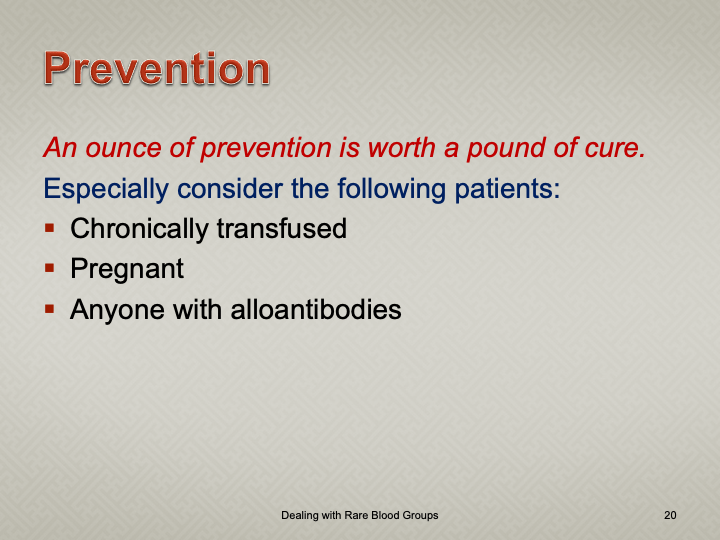
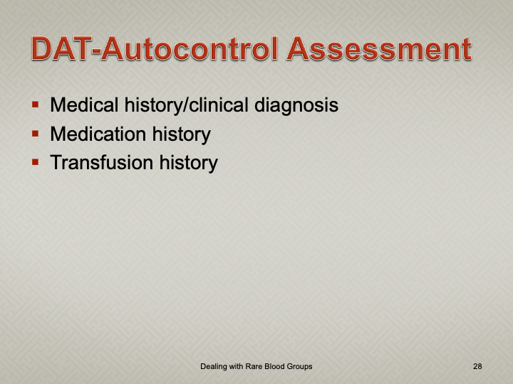
My practice across the globe has exposed me different rationales to performing antibody titration.  In my American training and practice (and also at international institutions following the American version of AABB accreditation), I only routinely performed titration of anti-D for Rh(D) hemolytic disease of the newborn and anti-A/anti-B for ABO-incompatible stem cell transplants AND ABO-incompatible renal transplants.

I have had heated arguments with some physicians who insisted they wanted titers for other antibodies.  The AABB Standards do not require this but leave it to the discretion of the Transfusion Service Medical Director.

In my entire career, I never worked in a blood bank or blood center which had optimal staffing or resources.  I focused on what was medically/technically necessary and even then still had shortages.  If performing a test does not change the clinical treatment, why perform it unless you are doing a research project!

Titration is a time-consuming, and until recently, a tedious manual task.  Recently some of the automated immunohematology analyzers offer a titration program.  We used the Ortho Vision Max which could perform both IgG and IgM titers within one hour—walk away!!  However, during that time, the titration procedure monopolized the analyzer.

Nowadays, low-anti-B-titer group A universal plasma and low-titer (anti-A and anti-B) group O whole blood may be offered as components.  At HMC Qatar, a preliminary study showed about 50% of units could be classified as low-titer (defined as a saline titer <1:128).  The amount of titration will require an automated analyzer.

The ABO-incompatible renal transplant program at HMC Qatar was modelled after Sweden’s Karolinska Institute.  However the latter site performed manual IgG and IgM titrations using Biorad/Diamed gels.

I did not have sufficient resources to commit staff to manual titration at HMC so I did a comparison study between the Ortho Max and the Biorad manual gel methods.  We were able to get good correlation and used the automated method for the transplant.

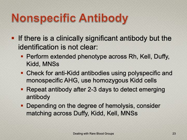
I still don not perform against performing titrations for antibodies other than anti-D.  I always ask, ‘Does the titration correlate with clinical severity?’  Unlike anti-D, antibodies such as anti-Kell and anti-c may be low titer but cause death.  Can anyone show me a definitive study that titers are useful except for transplants and Rh(D) hemolytic disease of the fetus/newborn?

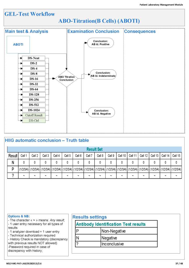
Since the method was working well on the Ortho equipment, I next established an interface to Medinfo.  The test was performed separately for IgG and IgM antibodies.  Medinfo recorded the reactions in all the wells.  The last well showing a 1+ reaction was interpreted as the titer (e.g. if 1:64 were the last 1+ reaction, then the titer was 64 in Medinfo).

The Medinfo process is shown below.

Building Your Blood Bank Software—Initial Considerations

In a series of posts, I will elaborate on how I built the processes and settings for a blood bank computer system in conjunction with the vendor’s software engineers.  This also applies to other laboratory software.

If you don’t know exactly what you are doing, how can you improve it?  Regardless whether you currently have laboratory software, you still need to optimize processes, determine critical control points, and plan improvements based on that.  A good manual system is the foundation for a good software build.

I was never taught in medical school how to do this.  I learned on-the-job at a time when software was quite rudimentary and mainly to record results.

Staffing:

For our first system, we used medical technologists to make settings for and administrate.  We thought that only those with a technical background in the field could do this.  It was moderately successful.  There was some antagonism between the technologist computer staff and the hospital computer department.  The technologists did not have a background in databases and programming;  the IT staff did not know the laboratory and were frustrated in dealing with the laboratory staff.

Later, to help reconciliate the two when a new hospital system was installed, we tried a different approach.  We found a database professional who was a very good listener.  Although he had no blood bank technical background, he could listen and map out the processes.  He was well-liked by the technologists who saw that he just wanted to understand their work and help them.  He was very successful in this endeavor.  I strongly recommend a software engineer as the lead in the project, one who can work with technical and medical staff to map out processes.

Unifying Processes Across Multiple Sites;

If your organization covers multiple sites, it is best to unify your processes as much as possible.  We built our dedicated blood bank system AFTER we had done this so the processes (except for some equipment differences) were the same everywhere.   This allowed us to move work between institutions quickly and makes system administration easy.

At one organization, I worked at, they had not done this.  They built their system based on the processes at the first site to go live, which was a small hospital with less than 10% of the workload.  It was not designed for the high-volume sites, and this was major problem as the larger sites were implemented. 

Capturing the Current State:

Most importantly, I cannot emphasize enough the need to capture the current state.  Take the time to do this properly and thoroughly.  This will help you whether or not you are building a computer system or just optimizing your manual processes.

At one institution in a non-blood bank system build, the administrative decision was to rush and not wait to complete this task so the actual processes were not captured—I actually rejected the proposed current state but was overruled.  The institution did not unify their processes as much as possible across sites.  The result was a suboptimal system that many/most people do not like:  should you blame the build or the limitations of the underlying software?  In my opinion, you can’t fully blame the software itself, if you didn’t design your build properly.

My Opinion: Use of Enzyme Panels

This is an updated version of a previous post.

Working for many years in the Middle East/Gulf, I have encountered significant antibodies that can only be detected at enzyme phase.  This is especially true of Rh system antibodies, particularly anti-c in an R1R1 patient.  I have attached an example.

The reasons I strongly recommend this practice are:

  1. Weak Rh system antibodies (as above)
  2. Confirmation of enzyme-labile antibodies, especially if there may be combinations of enzyme-labile and enzyme-resistant antibodies (e.g. anti-Fya and anti-c).

It is also important to consider which enzyme to use:  bromelin, ficin, or papain usually and sometimes trypsin or chymotrypsin.  They do not always attack at the same site.

In addition to most common MNSs and Duffy system antibodies, many Kell antibodies (e.g. K or K1, Kpa) are labile with papain:  however, with ficin they may be partially labile, unaffected, or even enhanced.

Using enzymes is a double-edged sword since they may enhance cold antibodies and thus cause nonspecific reactions.  Thus, I know many of you may not routinely include them in your workups.

It is essential to follow the manufacturer’s recommendations for their use.  If you make your own enzyme-treated cells and prolong the incubation, you may get false positivity.  You should also be careful about using potentiators with enzyme-treated cells—normally I run them in saline.

Since anti-c may cause severe hemolysis and severe hemolytic disease of the newborn, I am especially vigilant in my R1R1 patients, particularly females of child-bearing age and all chronically transfused patients.  I prophylactically match R1R1 patients with R1R1 RBCs in these categories, regardless if either anti-E or anti-c are expressed.  I have seen many examples where the anti-c is only detected at enzyme phase.

It is my practice to always include an enzyme panel.  I would be very interested to know your practices?  When do you use enzymes?

Investigation of Donor Unit Mislabelling

Principle:

All donor unit mislabeling is potentially life-threatening and must be stringently investigated as soon as possible after the discrepancy is detected.  Most importantly, if there is one error, there may be possibly ADDITIONAL donor unit errors (e.g. switch of donor tubes or units, etc.).  All donor units processed in the same batch must be also quarantined until the discrepancies are resolved.

Definitions:

Responsible blood bank physician:  specialist or consultant physician on-call at the time the discrepancy is detected

Policy Details:

The following steps MUST be performed as soon as possible:

  1. The Component Processing Supervisor or Senior Technologist must be IMMEDIATELY notified of any discrepancy.
  2. The Blood Bank Supervisor will inform the Division Head, Transfusion Medicine.  If the Head is not available, notify the Transfusion Medicine on-call.
  3. Quarantine ALL donor units collected and processed in the same batch.
  4. Obtain copies of all testing including photos of the gel/glass bead cards documenting the discrepancy.
  5. Obtain copies of all worksheets used in donor processing for the affected batch.
  6. Perform repeat ABO/D typing of ALL DONOR UNITS in the affected batch.  Any further discrepancies must be investigated and resolved.
  7. Identify all staff who were involved in handling the donor unit (phlebotomist, blood bank technicians processing and labelling the unit).  Identify those associated directly with the error.
  8. Submit all documents and photos to the Blood Bank Supervisor or designate.
  9. Prepare an occurrence/variation OVA report documenting all the data, findings, and interpretations.
  10. All investigations must be reviewed by the Supervisor, responsible blood bank physician, and one of the senior consultants.
  11. All such investigations must then be finally reviewed and approved by Head, Transfusion Medicine or his designate.  Only when the issue(s) are completely resolved and investigation is approved may the donor unit be properly relabeled and released into available stock.  Also, only at that time may the other units in the affected batch be released into available stock!!
  12. Photograph the correctly relabeled unit and attach it to the other documentation of the incident.
  13. If the discrepancy cannot be resolved, ALL units in the affected batch must be discarded.
  14.  The implicated staff’s personnel record should be reviewed for previous errors.   Appropriate disciplinary action should be taken and documented in the personnel record.  If a verbal warning is given, it should still be documented in the written record.
  15. If there is a systemic cause for the error, appropriate measures should be taken to minimize reoccurrence.
  16. All actions must be in accordance with the applicable regulations.

Order of the Steps in Serologic Testing

This is a teaching document I give to new staff, medical technology students, pathology and residents.  Very often I get the question, “Why can’t I just do the antiglobulin phase crossmatch first and then phenotype the RBC unit?” Or:  “Why do I have to add reagents in a particular order?”

My practice has always been to select an antigen-negative RBC unit first, then do the antiglobulin-phase AHG crossmatch.  This way I know that the unit was definitely phenotyped before release.  Likewise, the blood bank computer now only offers antigen-negative units for allocation and then crossmatching if there is a clinically significant antibody.

In a manual setting without a blood bank computer system, performing the AHG crossmatch may yield a negative result, even if the unit is antigen-positive.  With storage, some antigenic expression is weakened so it may not be detected at the time of crossmatch.  Yet, there may still be enough antigen present to cause hemolysis.  Not detected does not necessarily mean not present!!

I expect that many inexperienced staff may be tempted to forego the antigen typing if the AHG crossmatch is negative.

This is an analogous logic to the question, “Do I add the cells or the antiserum/plasma/serum first for the reaction?”  If you add the cells first, you may forget to add the patient’s plasma/serum or a typing antiserum and you might not be able to detect the omission by looking at the tube or gel.  Actually, I once recommended to one vendor that it color the typing antiserum so it was conspicuously showing on the gel.

I was taught that this is a matter of discipline to ensure that all steps are performed.

However, for every practice, there has to be flexibility.  If there is no typing reagent or if it is very expensive or in short supply, one may have no choice but to do the AHG crossmatching first.  Often there is still another option:  one can often preliminarily screen units first before using a rare reagent—examples:

  • Check if patient is group O first and antibody screen panreactive in suspected anti-H.
  • Check P1 typing first if there is a suspected anti-PP1Pk.
  • Check the antibody screen for panreactivity first for antibodies of high-incidence or prevalence antigens.

ABO Incompatible Stem Cell Transplant: What ABO type of RBCs Should I Use?

In an ABO-incompatible stem cell transplant, both donor and recipient RBC types may be present.  Likewise, immune effector cells from both the donor and recipient may be present.

Using group O RBCs and AB plasma is an option but there are limited supplies of both.  Since we use RBCs in additive solution (SAGM), only minimal residual donor plasma is available and unlikely to be clinically significant.

Here is my approach:

  1. Use fresh specimen to perform forward and reverse type
  2. Check the reverse type:  does it show either anti-A and/or anti-?
    1. If anti-A detected, do not transfuse group A RBCs.
    2. If anti-B detected, do not transfuse group B RBCs.
    3. If both anti-A and anti-B detected, continue using only group O RBCs.

Massive Transfusion: When to Revert to the Original ABO Type

In a massive transfusion setting, the patient’s forward and reverse type will reflect the use of group O RBCs and the patient may even fully type as group O, depending on the number of units transfused.  Group O RBCs are very precious and in short supply so we need to switch the patient back to his/her own type as soon as feasible.  RBCs in additive solution (e.g. SAGM) have only minimal residual plasma so the load of anti-A,B from the O cells is minimal.  In my organization, we did not titer ABO hemolysins in blood donors.

Here is my approach:

  1. Use fresh specimen to perform forward and reverse type
  2. Check the reverse type:  does it show either anti-A and/or anti-B from the group O massive transfusion?
    1. If anti-A detected, do not give group A RBCs.
    2. If anti-B detected, do not give group B RBCs.
    3. If both anti-A and anti-B detected, continue using only group O RBCs.

Some people would recommend performing a full AHG crossmatch using the patient’s current plasma and RBCs of the original ABO type:

  1. If compatible, return to the original ABO type.
  2. Otherwise, continue using group O RBCs.

I did NOT do the full AHG crossmatch and had no hemolytic transfusion reactions if the RBCs were compatible with the current reverse type plasma.