Transfusion Medicine, Blood Bank IT, Pathogen Inactivation, Plasma Fractionation, COVID-19 Convalescent Plasma Production
Category: Patient
Includes compatibility testing (AHG, electronic, immediate-spin), antigen typing, antibody screening and identification, direct antiglobulin testing, elution, transfusion reaction and drug reaction workups, component typing tests upon receipt in hospital blood bank, release and return of components
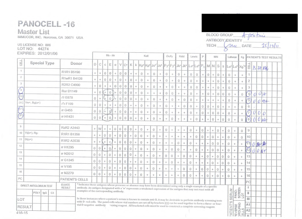
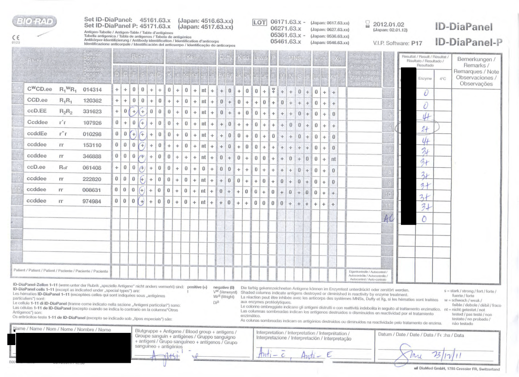
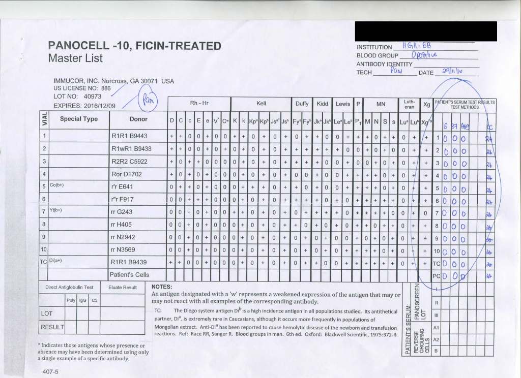
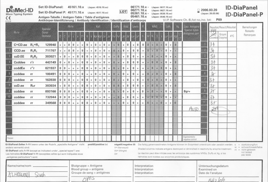
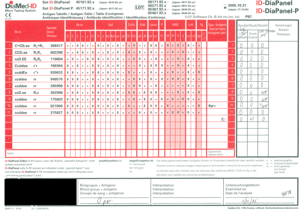
Working for many years in the Middle East/Gulf, I have encountered significant antibodies that can only be detected at enzyme phase. This is especially true of Rh system antibodies, particularly anti-c in an R1R1 patient. I have attached an example.
The reasons I strongly recommend this practice are:
Weak Rh system antibodies (as above)
Confirmation of enzyme-labile antibodies, especially if there may be both enzyme-labile and enzyme-resistant antibodies
It is also important to consider which enzyme to use: bromelin, ficin, or papain usually and sometimes trypsin or chymotrypsin. They do not always attack at the same site.
In addition to most common MNSs and Duffy system antibodies, many Kell antibodies (e.g. K or K1, Kpa) are labile with papain but less so or not at all with ficin.
Using enzymes is a double-edged sword since they may enhance cold antibodies and thus cause nonspecific reactions. Thus, I know many of you may not routinely include them in your workups.
It is essential to follow the manufacturer’s recommendations for their use. If you make your own enzyme-treated cells and prolong the incubation, you may get false positivity. You should also be careful about using potentiators with enzyme-treated cells—normally I run them in saline, not LISS!
Since anti-c may cause severe hemolysis and severe hemolytic disease of the newborn, I am especially vigilant in my R1R1 patients, particularly females of child-bearing age and all chronically transfused patients. I prophylactically match R1R1 patients with R1R1 RBCs in these categories, regardless if either anti-E or anti-c are expressed.
I would be very interested to know your practices? When do you use enzymes?
Anyone reviewing antibody panels, especially in the Middle East/Gulf region, encounters many panels for which no antibody specificity is identified. As a transfusion medicine physician who often got called during the night for release of RBCs for patients with “nonspecific” pattern, this was a big headache.
Is it “nonspecific” because there isn’t a clinically significant antibody OR the technologist did not perform the testing or its interpretation correctly? Does it need further testing? Do I release blood components at this time?
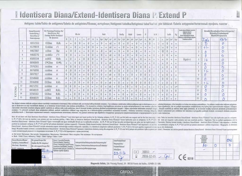
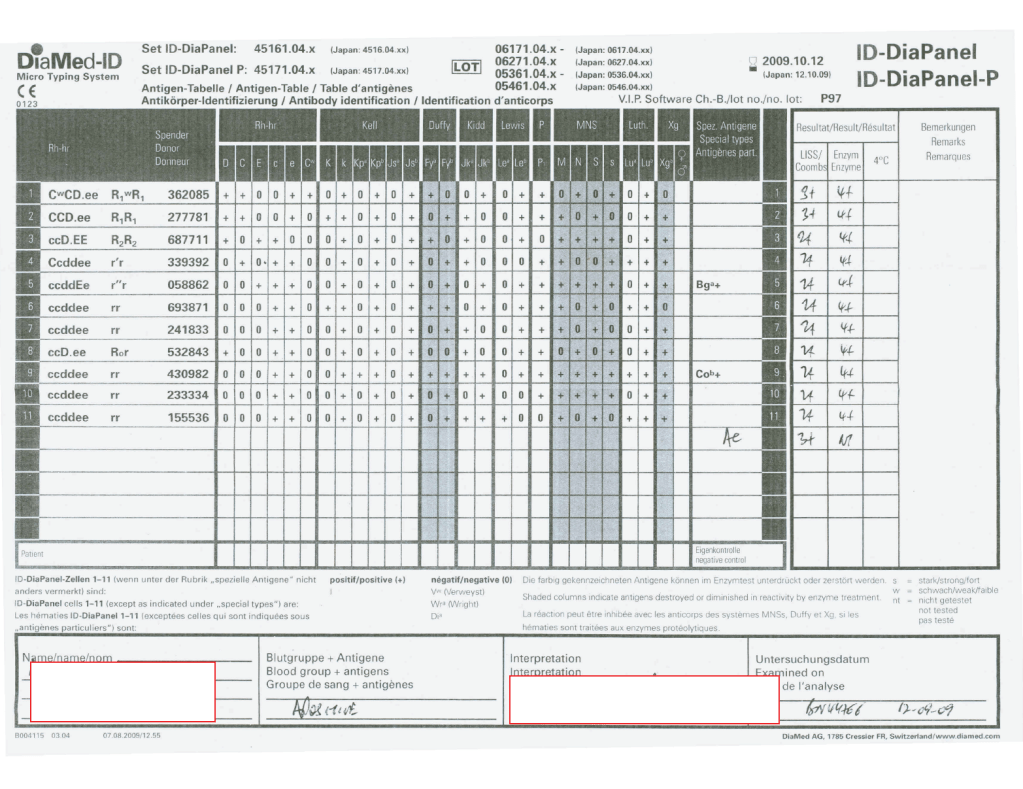
I have attached an example of an antibody panel that has a few weak to 1+ reactions using a gamma-whole-molecule IgG AHG reagent. Notice that they are enzyme-labile.
I have worked in this region both before and after gel/glass bead technology was introduced. In the good old days, I formerly only used a gamma heavy-chain specific AHG in tubes with LISS. Nonspecific reactions were few.
When we adopted gel and at some sites glass bead columns, there was only a choice between polyspecific AHG and whole-molecule IgG AHG. The rate of non-specificity soared from less than 10% to over 30% of panels. The reactions were reproducible over multiple technologists at multiple sites.
When I repeated the same specimen against a manual tube panel using gamma-heavy-chain specific AHG, the reactions disappeared in the majority of the cases. Why?
Heavy chain specific reagents do not detect light chains. Light chains are the same in IgM and IgG antibodies. Both polyspecific and whole molecule IgG AHG detect light chains. Thus, high-thermal-amplitude cold antibodies may be detected with the latter but are unlikely to react with heavy chain specific reagents.
When I used manual tube reagents, I would have missed these reactions, but I do not recall this having clinical consequences except in very rare circumstances (refer to my previous post about the anti-Jka only detectable with polyspecific reagents—I have seen 3 in 30 years).
I have asked various gel/glass bead manufacturers to provide me with heavy-chain gamma-specific AHG, but none have agreed.
Normally, for automation, I use IgG-whole molecule AHG. If the reactions are nonspecific, we repeat the testing manually using the gamma-heavy-chain-specific AHG. If negative, I ignore the nonspecific reactions and use the same gamma-heavy-chain-specific AHG for full antiglobulin-phase crossmatching.
In general, with nonspecific reactions, I recommend the following:
Always do enzyme panels, sometimes with both papain and ficin reagents: many Rh antibodies are optimally detected only at enzyme (example: R1R1 with apparent anti-E at AHG but the anti-c only seen at enzyme)
Perform an extended Rh/Kell/Duffy/Kidd/Kell/MNSs/P1 phenotype and specifically check for those negative typing results AND for dosage (could the antibody only be detected in homozygous cells like many anti-M are?).
Perform classical room temperature, 37C, and finally AHG phase testing. Routinely I do not do this since antibodies not detected at 37C are unlikely to be clinically significant. Sometimes, the AHG phase reactivity is a cold antibody of high-thermal amplitude.
If the Jka or Jkb antigen typings are negative, repeat using a polyspecific AHG reagent.
Use additional panels from multiple manufacturers. Some reagents detect more nonspecific reactions than others.
Try other potentiators than LISS such as PEG.
Check the outdate of the panel and reagents: if less than 1 week remaining, consider repeating with fresh reagents and getting a new patient sample.
Finally, if you still cannot define the specificity, consider repeating after several days. Maybe it is a newly emerging or an anamnestic response.
I emphasize as a physician, I do not care to see all possible antibodies present in the specimen but rather only those likely to be clinically significant.
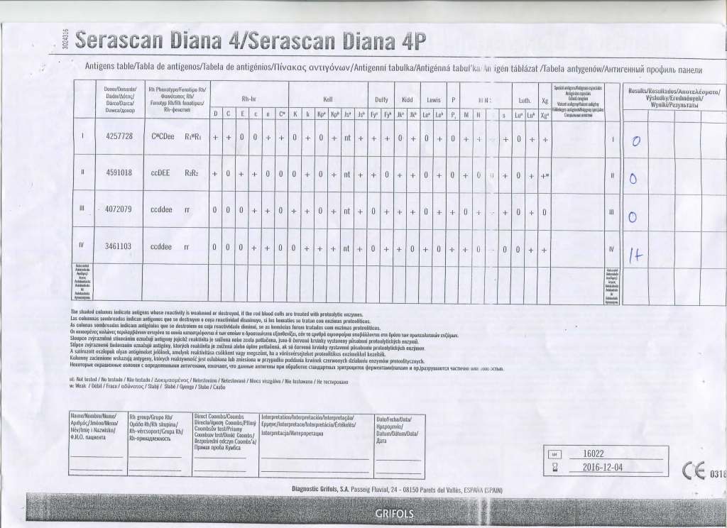
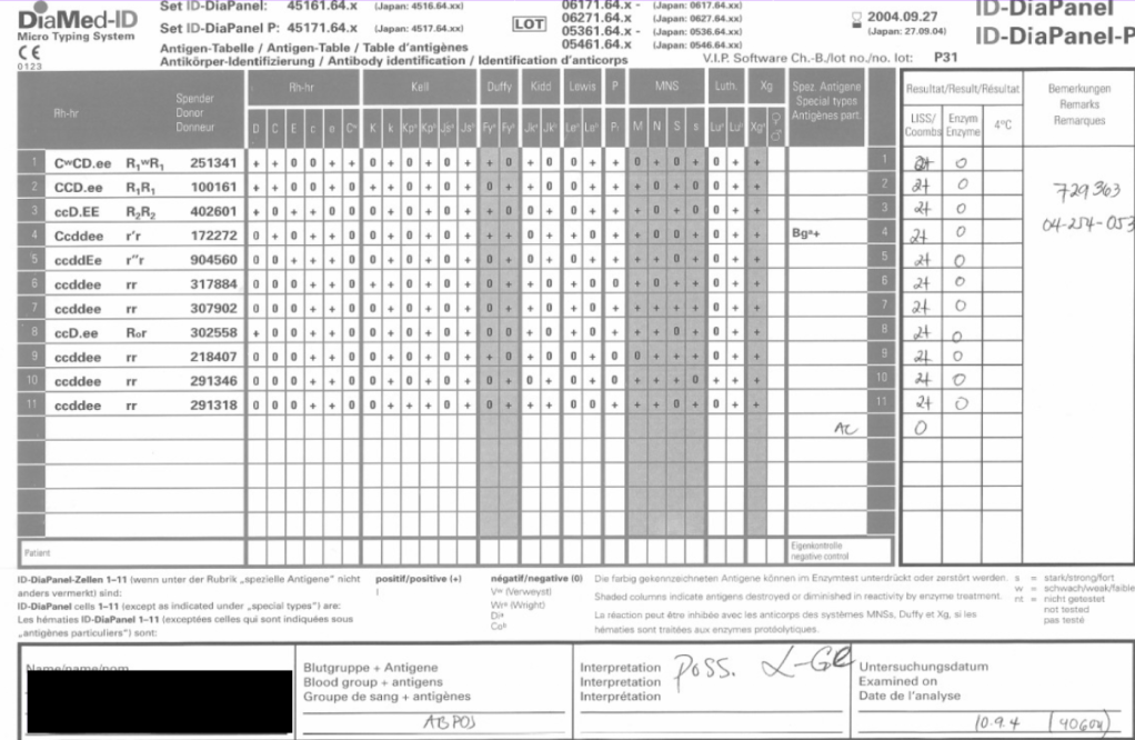
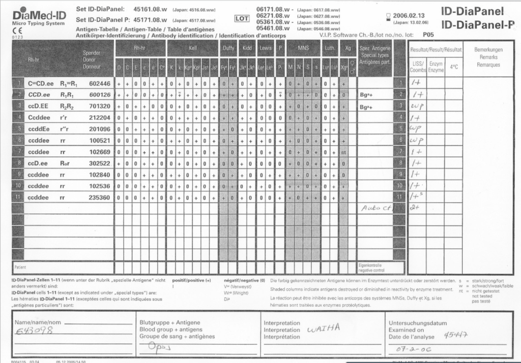
As a Transfusion Medicine physician, I reviewed all antibody workups. Here are three different panels, all appearing to be panreactive, at AHG phase. Note the differences:
Autocontrol is positive, enzyme is panreactive and enhanced
Autocontrol is negative, enzyme phase shows no reactions
Autocontrol is negative, enzyme phase is panreactive enhanced
In the middle of the night, if I am called to select blood, I always keep these patterns first and foremost in my mind.
Case 1:
If the autocontrol is positive at about the same strength as the panreactivity. I can state that this is probably a warm autoimmune pattern WAIHA (although I cannot rule out underlying clinically significant alloantibodies obscured by this pattern and need to do autologous auto-absorption ZZAP). Both warm autoimmune antibodies and certain drug reactions may cause this pattern. Use least-incompatible crossmatch matching any significant specificities you found by ZZAP and observe the patient closely throughout transfusion.
Case 2:
If the autocontrol is negative and all reactions are enzyme-labile—and if you live in the Middle East/Gulf region, then you have a presumptive anti-Ge2. You can safely ignore this antibody and release least-incompatible crossmatch RBCs, regardless of the strength of the reactions.
Case 3:
If the autocontrol is negative and the reactions are unchanged or enhanced by enzyme, BE AFRAID, VERY AFRAID!! This is an antibody to a high-incidence antigen. These can be very dangerous. In the Middle East/Gulf region, consider anti-H, anti-k (cellano), anti-Kpb, anti-PP1Pk, and rare antibodies to the MNSs such as anti-U or other MN system deletions. There are many other possibilities, e.g. anti-Fy3.
You need to perform extended antigen typing across the major Rh antigen, Kell, Duffy, Kidd, MNSs, P systems (at least P1):
Run H lectin to rule out Bombay Oh or Parabombay.
Some Rh system deletions and Rh null show pan-Rh reactivity—check D, C, c, E, e typings.
Anti-k (cellano) will be suggested by k-negative phenotype.
Unusual, weak or absent reactions with M and N reagents suggest something like En(a)-negative or similar.
Absent P1 with no other findings, you must rule out anti-Tja (anti-PP1Pk).
I have seen all of these specificities during my time in the Middle East. All of these antibodies can be clinically significant and often life-threatening.
Conclusion: NEVER NEGLECT TO REVIEW THE AUTOCONTROL!!!
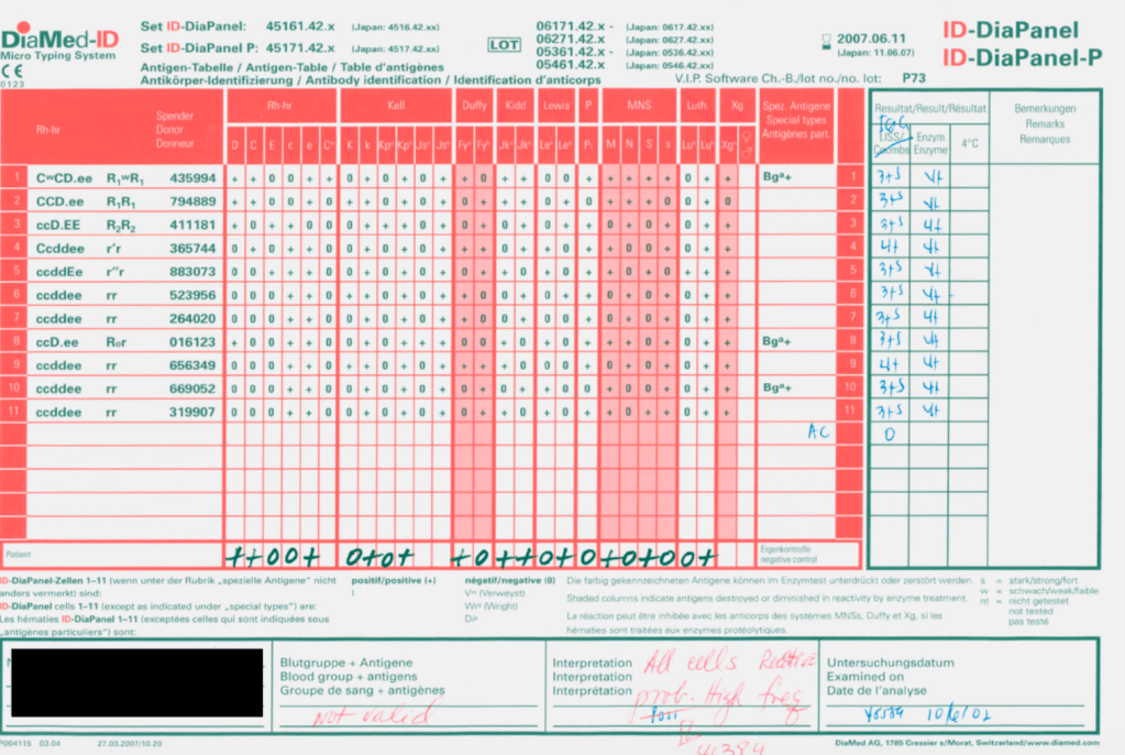
Whenever I had a “nonspecific” antibody, I had to first rule out issues with the reagents themselves. The following example shows weak to 2+ reactions in the panel cells and autocontrol.
The variability in the reactions made me initially uncomfortable about called this WAIHA. I then checked the panel details: the testing was done only six days before the panel outdate.
I told my staff to repeat the workup with the new panel expiring five weeks later. The difference is astounding!!
Remember: if you work in the Middle East, the environmental conditions can be extreme in summer (>50C). Do you know how your reagents were handling during transport?
My advice: if you are concerned there is a clinically significant antibody but cannot discern it, consider repeating the workup using fresh reagents.
Blood Bank instruments may perform tests and release test results in a numerical or alphanumeric format or both. For example, nucleic acid and enzyme immunoassay may release a qualitative result (e.g. positive, reactive, borderline/grayzone, negative, nonreactive). Alternatively, the machine may release the signal to cutoff ratio (S/CO) as a numeric result.
Blood bank software may use either kind of result on which to base interpretative rules for acceptability of the donor. The qualitative result criteria are based on the quantitative SC/O but the equipment automatically interprets this. The S/CO ratio of 1 is the cut-off point. Thus a value of 0.99 is negative and the value 1.01 is positive. But is it really so clear-cut since the difference between the two is so small? Thus, some people have added the term grayzone for values close to but below the cutoff. Could a value of 0.95 be an early infection?
I personally prefer to see the actual cutoff but use the manufacturer’s criteria for interpretation. As a physician, it is good to review the S/CO on serial exams. If a borderline or grayzone result becomes positive, then perhaps the original result indicated early infection. The question still remains, what is the grayzone? 0.95 to 0.99, 0.90 to 0.99, etc. Some accrediting schema have not used grayzone for interpretation.
With Medinfo’s blood bank software, I could chose either option or both—or at least store the S/CO as a nonreported result for subsequent review. I could even chose, test by test, in a series between reporting either S/CO or the qualitative result.
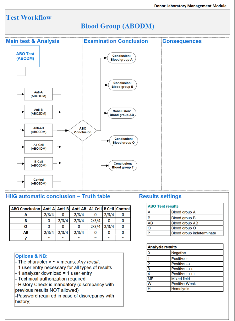
Semiquantitative results, e.g. in {0, 1+, 2+, 3+, 4+} are qualitative and could also include mixed field (mf) and hemolyzed (h). I showed examples of this with ABO/D antigen typing in a previous post—see attachment.
On the contrary, the results from blood production equipment may include parameters such as time of preparation, original volume, final volumes for each component, platelet yield index as an indirect measure of platelet count. When there is pooling, the final total volume is critical to determine if pathogen-inactivation procedures and platelet additive solution can be used. This is a much more complicated interface.
The blood production equipment interface issues will be considered in a future post.
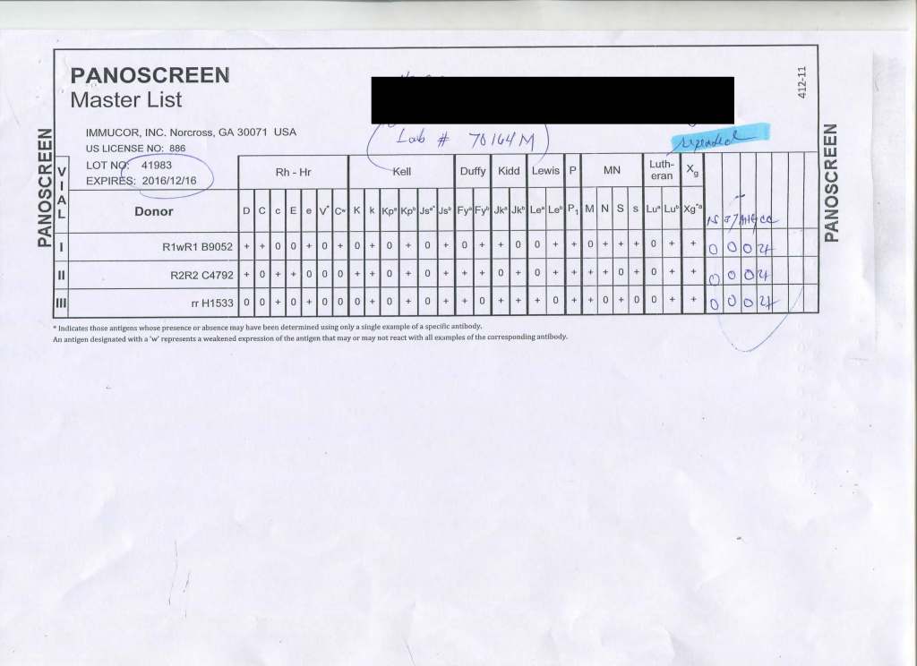
This is a rare anti-Jka that only reacts with polyspecific AHG and only in Jka+ homozygous reagent cells. In the past three decades in the Middle East, I have only seen three of these.
Almost all anti-Jka and anti-Jkb antibodies in my experience can be detected using monospecific, gamma-heavy-chain AHG reagents (I personally prefer this latter reagent to avoid cold antibody interference.)
If you have a nonspecific antibody, never discount this possibility and then run polyspecific reagents and check for dosage. I also recommend extended antigen typing (e.g. Diamed/Biorad Profile Cards 1-2-3).
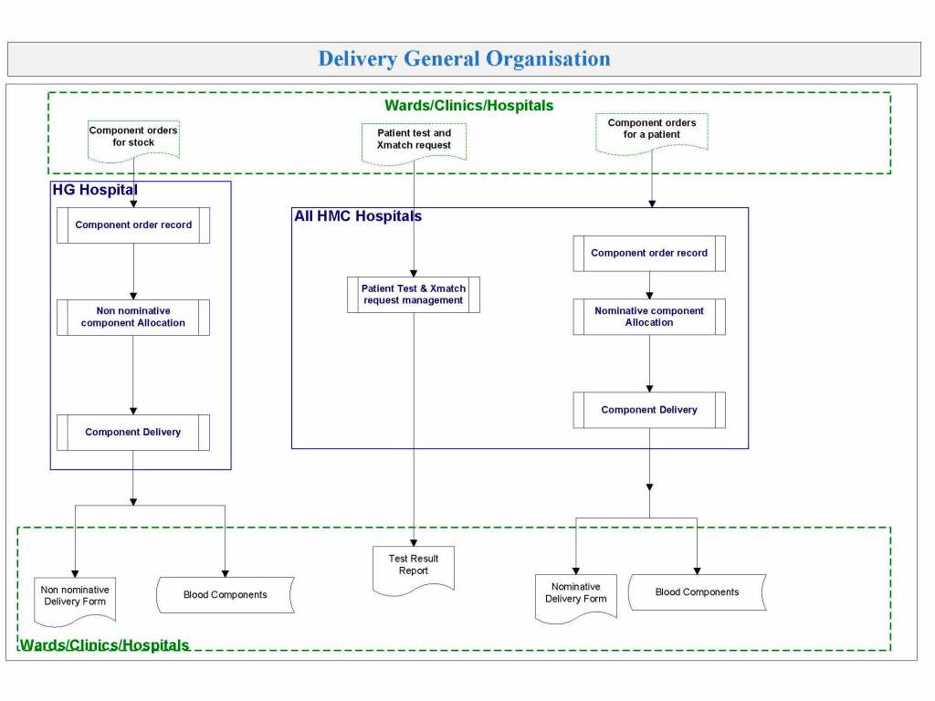
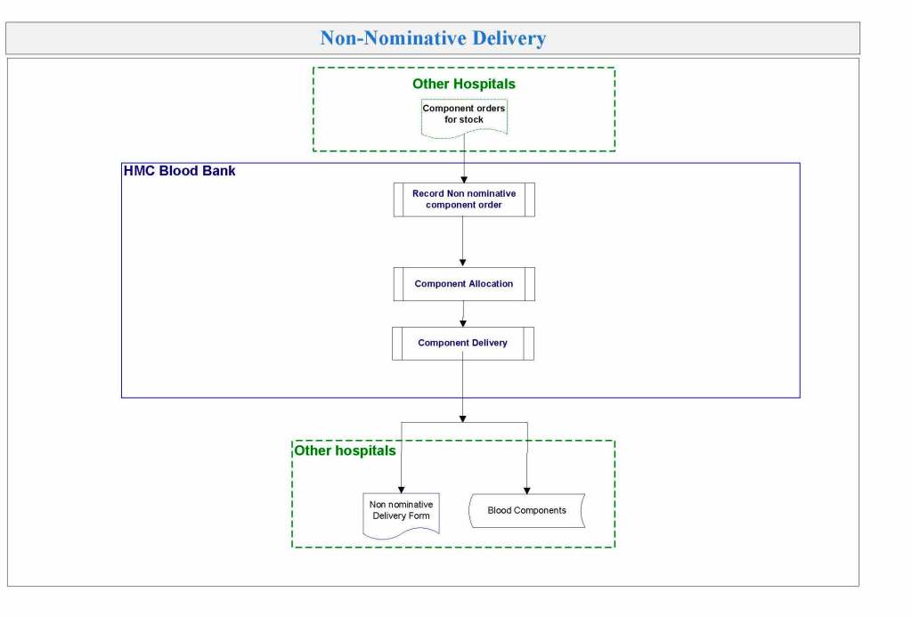
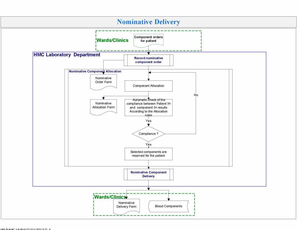
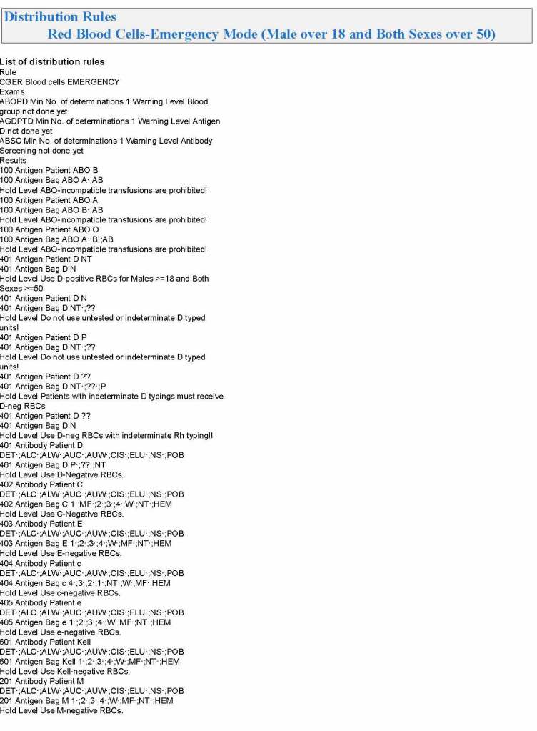
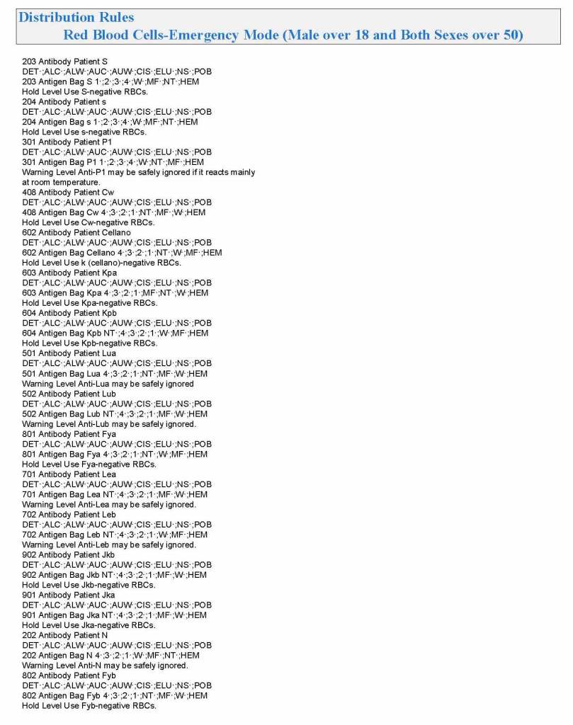
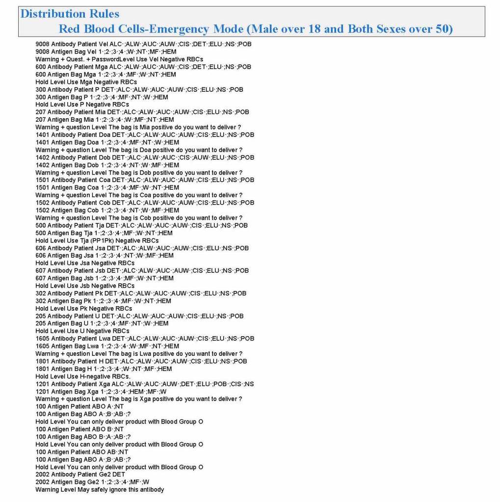
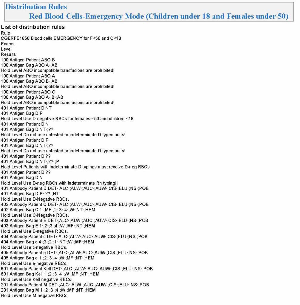
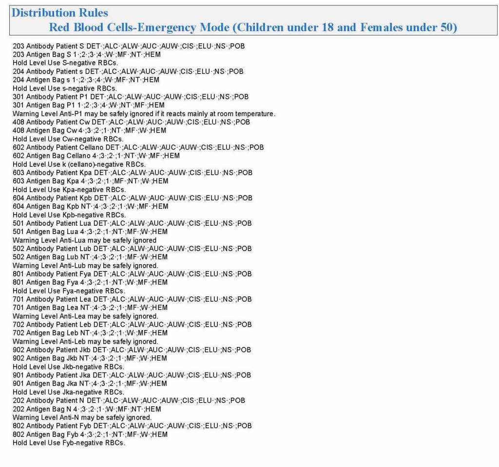
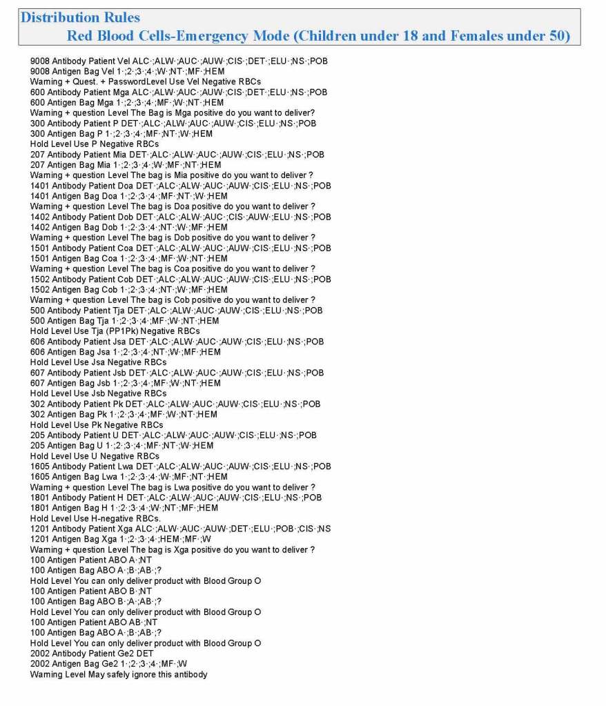
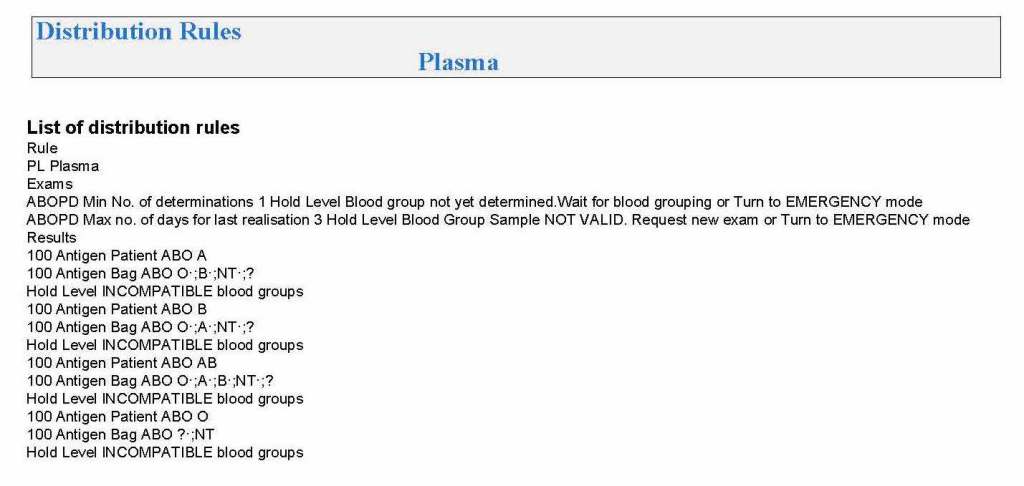
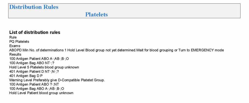
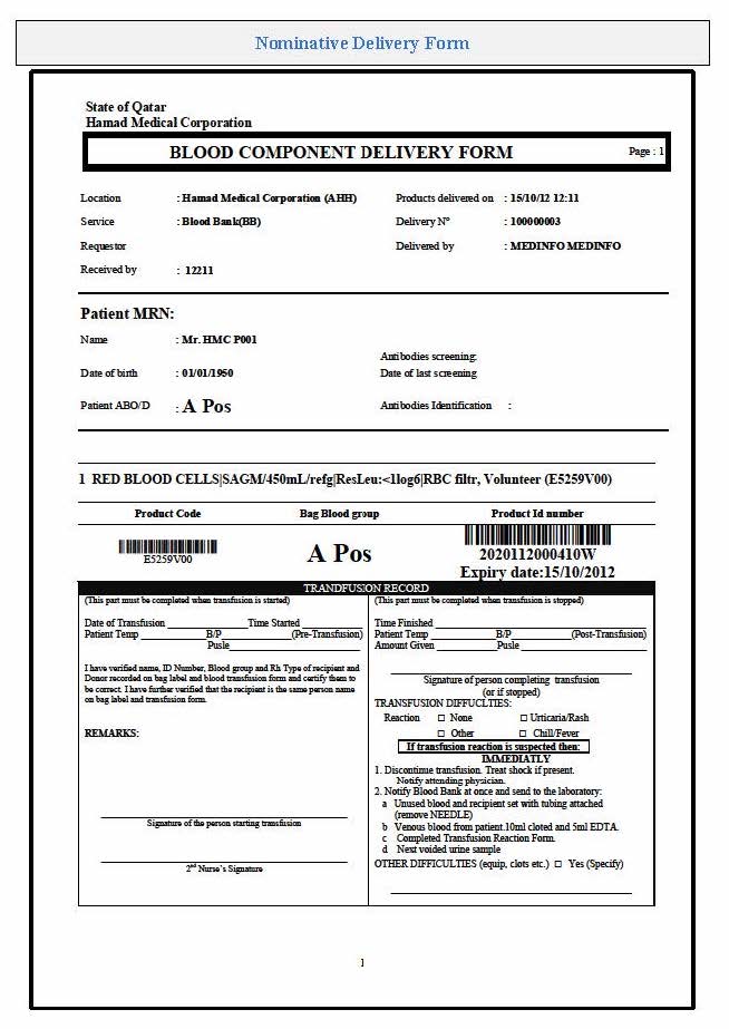
In my recent post, I provided sample flows and parameter mapping for delivery of blood components. The final components from the component preparation center may be sent to various depots (freestanding location and/or hospital blood banks. There should be complete traceability for every step (from donor reception, collection, testing, and processing) transport between locations, and finally the exact storage site, which might include which refrigerator/freezer/incubator and even shelf/position number for each component is stored. The end of that document showed rules for type/antigen matching.
For disaster planning, rapid inventory enumeration by type is very important. This can be very time-consuming manually. With our Hematos blood bank system, we could quickly get total inventory across the Qatar or by hospital in less than one minute. We could also quickly find antigen-matched units across the system and reserve it at any one site for another if necessary.
Smart blood bank dispensing refrigerators, as offered by Haemonetics and Angelatoni, may also serve as depots and take the place of a hospital blood bank for some dispensing. These solutions can also capture vital information about the storage conditions of the components and prevent release if the storage criteria are not met. They can also interface with blood bank computer systems and use the main system’s logic for the dispensation rules.
Upon receipt at the hospitals from the blood processing center, the forward ABO and D typing must be confirmed. We used D reagents which detected partial D so that we would call such donor units as D-positive. However, if a patient type reagent insensitive to partial D types were used, it is possible for a unit to be typed as D-negative whereas in the donor center it might be D-positive. Sometimes, nothing types consistently as D-positive: all you can say is that with a particular reagent and lot number, there is or isn’t reactivity.
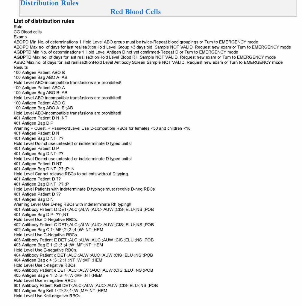
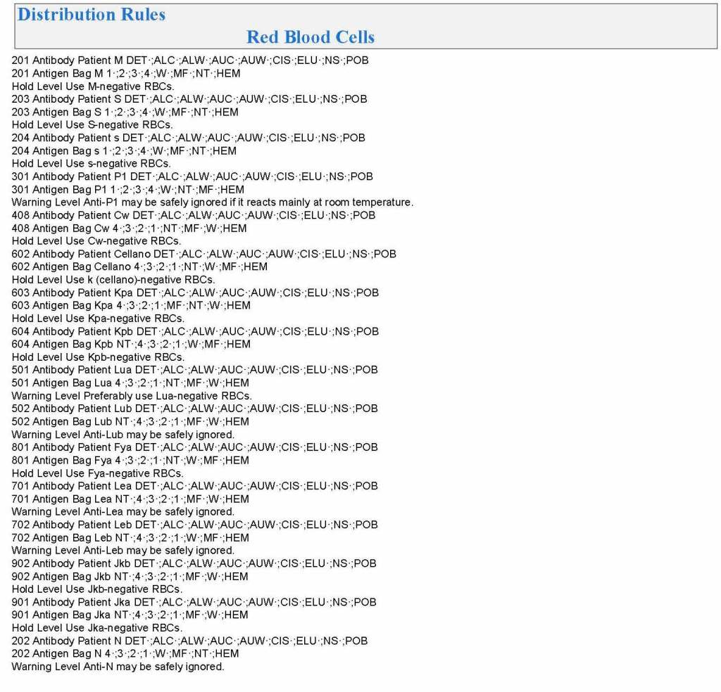
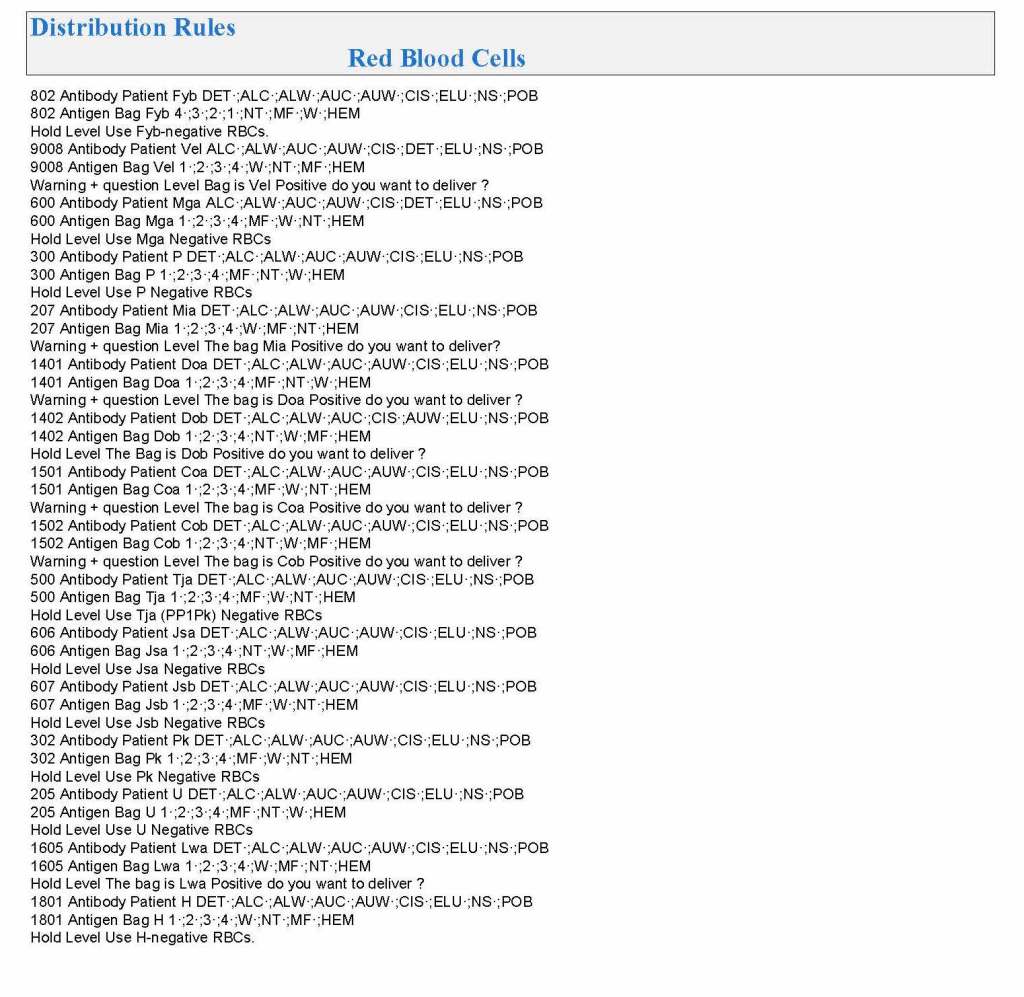
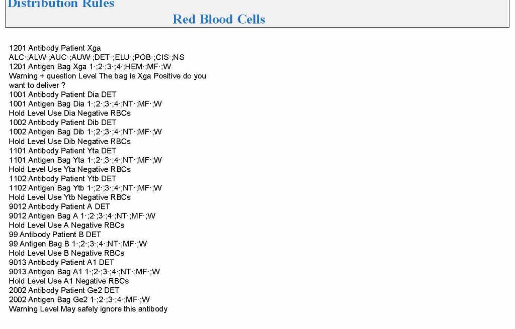
The greatest complexity is for RBCs since potentially so many antigens exist. Criteria for matching/ignoring certain antigens must be made. Critically significant antibodies such as the Kell, Duffy, Kidd, and certain Rh (D and c) must be antigen matched. A robust blood bank computer system can enforce these rules.
For other components, antigen/typing may be less important. In fact, in most situations, any type of platelets can be given to anyone (except neonates). Despite the potentially incompatible plasma, there is rarely significant hemolysis. In fact, if pooling platelets without regard to blood types is done, a platelet transfusion is a common cause of a positive direct antiglobulin test DAT—something that is not clinically significant. No one died of a positive DAT by itself for this reason.
Specific rules for compatible plasma types are important, but nowadays, low-titer group A plasma may be used like universal AB plasma. The challenge is to be able to perform the ABO titration (specifically anti-B) quickly—titration can be a slow process, even with automated equipment. A similar situation for low-titer, universal group O whole blood requires both anti-A and anti-B titration (I will return to this topic in a future post).
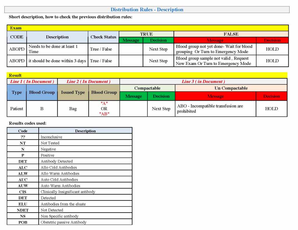
In the previous post, the Medinfo document for Inter-Depot transfer had many pages of rules for matching RBC antigens. Multiple actions were available:
Forced Match, no release of untested or antigen-mismatch (e.g. anti-Kell requires specifically tested K-negative RBCs).
Match Optional (e.g. anti-Lea/Leb does not require Lewis-matched blood): system would flag a comment showing the antibody specificity, user would respond Y/N, and select units
Least-Incompatible (e.g. WAIHA): requires sufficient privilege (senior technical or transfusion physician) to authorize release
Permitted Incompatible (e.g. give C-positive to patient with anti-C in times of shortage or complex multiple antibodies without fully matched blood available): requires sufficient privilege to release
Fully Allowed (e.g. group O RBCs to group A, AB, B patients)—no flagging, allocation of units permitted
Prohibited Under Any Circumstance—NO Override Permitted (e.g. group O for Bombay Oh, c-positive for anti-c, K-positive for anti-K)
To avoid mistakes, the blood bank computer system enforced the rules. There was no mercy. Only specific individuals could override this( in many cases, but certain allocations (e.g. group O RBCs to a Bombay Oh patient) were not permitted under any circumstances.
Prophylactic Antigen Matching:
Please also refer to my prophylactic antigen matching post made last week for the rules I selected for Qatar.
Prophylactic antigen matching is common in Europe. I have been doing this during the many years I have worked in the Middle East. Most patients were not local nationals but transient. They would return to their home countries where blood bank testing (antibody screening/identification/antigen matching) or intrauterine exchanges might not be always available.
For pregnant patients, we would prophylactically match K-negative and c-negative—regardless if there were antibodies detected—R1R1 units for R1R1 patients. At the end of my time in Qatar, we had several pregnant women with various Rh deletions, so we added routine extended Rh(D) and Kell typing to all.
For sickle cell patients, especially African type, I would prophylactically match Rh antigens (D, C, c, E, e) and Kell because of the polymorphisms in the CE gene, some of which may lead to pan-Rh antibodies.
I would consider selective prophylactic antigen matching in chronically transfused populations, again regardless if clinically significant antibodies were detected.
If a patient makes any antibody, regardless if is clinically significant or not, I would consider that patient as a candidate for prophylactic antigen matching (but NOT necessarily for a clinically insignificant antibody).
In Qatar, blood bank services (testing and components) were not charged to the patient. In many other parts of the world, blood bank is a cost center. No prophylactic antigen matching may be routinely performed. If it is done, it must be charged to the patient or the hospital must assume the cost. I have gone to conferences in such locales where not even R1R1 patients were not matched and subsequently developed anti-c, which complicated management. It would have been cheaper to do the antigen matching than to pay for the consequences of the alloimmunization.
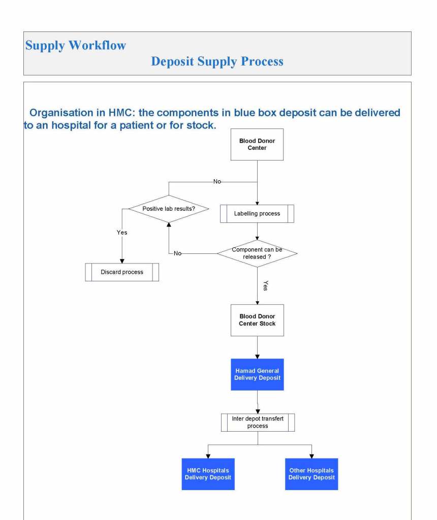
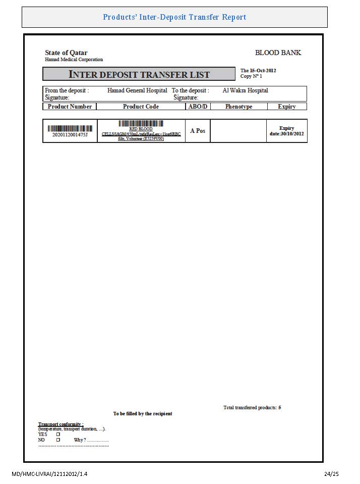
While I was working in Qatar, this was the overall process for transfer and allocation of blood components. Once they were finally labelled (only possible if all criteria had been meet), they were transferred from the Blood Donor Center BDC to Hamad General Hospital Blood Bank, from which they were distributed to all hospital blood banks in Qatar. Similarly, units could be transferred between the various hospital blood banks.
One could track components as being in:
BDC
Transit BDC to HGHBB
HGHBB inventory
Transit HGHBB to another hospital blood bank
Transit between any two hospital blood banks
An inventory manifest would be printed to show all transferred units.
For patient use, allocation rules applied which would determine if an electronic or a full antiglobulin-phase crossmatch could be used and whether specific antigen-matched components were required.
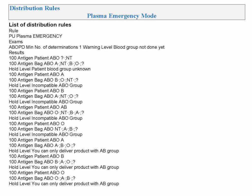
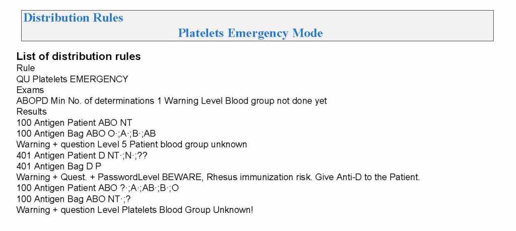
There were also separate rules for emergency release if the standard criteria could not be met.
Replacement fluids and volume (e.g. normal saline, 5% albumin, ACD-A, blood components, solvent-detergent treated plasma SDP)
Calcium replacement (calcium gluconate IV)
Orders for laboratory testing prior to and after the apheresis (usually CBC, PT, APTT, fibrinogen, total protein, albumin, calcium, LDH for TTP/HUS)
Apheresis nurse convey the order to the ward/clinic.
The requesting clinical service will place the order in the hospital information system.
If a verbal order is made, TMS physician will sign his verbal orders, a copy of which will kept in the Apheresis Unit.
If the hospital computer system is used, the TM physician will enter his orders and the apheresis nurses will take off the orders electronically.
If a stem cell product is collected:
TM will label it with a unique alphanumeric identifier barcode, i.e. an ISBT specimen label with check-digit that is generated by the Medinfo Hematos IIG system .
Cellular Therapy Laboratory staff in conjunction with the apheresis nurse will sign-out the collection from TM to CTL using two donor identifiers: patient name and the unique alphanumeric sequence barcode (same as process of sign-out of blood component).
Note: Use of Medinfo HIIG for stem cell collection is out of scope of TM.
Cellular Therapy Laboratory:
Directly receive collected product from apheresis nurses will use the procedure outlined in 7.2 above.
Most Responsible Physician:
Initiates request for apheresis with Division Head. Transfusion Medicine or covering Transfusion Medicine physician.
Obtains informed consent from patient
Arranges for proper venous access (rigid-bore central line or AV shunt)
Is responsible for clinical support during the procedure
Provides medication changes (e.g., substitution of non-ACE-inhibitors for hypertension therapy) as requested by the Head, TMS or Transfusion Medicine Physician
Ward/Clinic Nursing Responsibilities:
Ward/clinic nursing staff effect written orders to obtain replacement fluids (albumin, blood components, crystalloid), calcium, and order tests in hospital computer.
Ward nurses provide support (e.g., obtain medications, blood components, and miscellaneous items such as gauze, etc. and bring them to patient’s room) DURING THE PROCEDURE.
As per Apheresis Policy (TRM-PM-410-000-000-01), the assigned staff nurse from the unit shall contact the Blood Donation Center.
The assigned clinical staff nurse from the unit should call the local Blood Bank to request preparation of the requested components, e.g. RBCs, FFP, or cryoprecipitate and follow up.
To avoid unnecessary delays before calling the Blood Donor Center Apheresis Staff Nurse the assigned unit staff nurse shall inform the Blood Donation Center Charge Nurse when the following have been completed and are available on the ward:
Signed, informed consent
A central line with a large boredouble lumen.
Completed blood laboratory results
Only results within 24 hours of the intended procedure may be used to write the order.
In some cases, more recent results may be required (e.g. TTP) at the discretion of the Transfusion Medicine physician.
The ordered replacement fluids such as normal saline, thawed or liquid plasma, cryo-poor plasma, albumin and red cell units available in the ward
Calcium gluconate (ordered amount in 100 ml normal saline) available
The assigned apheresis nurse shall then perform the required therapeutic apheresis procedure strictly following the related TRM standard operating procedure.
Note:
Apheresis is a SCHEDULED, high-priority procedure like other invasive procedures. The ward must make certain that the patient is available for the procedure at the scheduled time and place.
Normally, apheresis procedures are performed 0600 to 1500 hours daily.
The service is ONLY available 24/7 for emergency procedures outside this time frame. Emergencies must be approved by the TM physician.
Transfusion Medicine does not guarantee that non-emergency procedures will be performed outside the 0600-1500 time frame.
The most-responsible physician MRP is responsible for the patient’s concern form, establishing the proper venous access (rigid-bore central line), and providing coverage ON-SITE during the procedure. The apheresis nurse may not leave the patient’s bedside during the procedure.
The apheresis nurse reports directly to the Division Head, Transfusion Medicine or the covering TM physician, not to the ward or MRP. Any issues about changing the apheresis orders must be approved by the Head, TM or the covering TM physician.
All other procedures must be cancelled during apheresis.
If the patient is not available at the specified, scheduled time, the procedure will be cancelled.
If ward or MRP support is not available, the procedure will be cancelled.