Teaching Document: Validation Process

This is a teaching document for medical technology and transfusion fellows to explain the general structure of a validation.

Principle:

All validations must be planned.  A validation protocol must be prepared with specific criteria for acceptance.  All validations with attached evidence must approved by the Head, Transfusion Medicine.

Policy:

  1. A written validation protocol must be prepared in the advance and at least including the following:
    1. Specific parameters and number of iterations to be performed
    1. Designated staff to perform validation
    1. Documentary evidence of the testing
    1. Specific acceptability criteria
  2. The completed validation protocol must be submitted to the Division Head, Transfusion Medicine, or designee for review.
  3. Once the validation plan has been reviewed, it must be performed by the designated staff.
    1. Software validations will be performed in a specific test environment, not in the live, production system.
  4. The completed validation document, including screenshots of the software functionality if applicable, must be submitted to the Division Head, Transfusion Medicine for review.
  5. The equipment or software may only be used if the acceptability are met AND the validation is approved by the Division Head, Transfusion Medicine or designee.
  6. The completed validation protocol will be stored in the document control system.

Reference:

Standards for Blood Banks and Transfusion Services, Current Edition, Bethesda, MD, USA

Blood Component Variances

Principle:

AABB Standards requires that all variances are documented and investigated and corrective actions taken when necessary.  Any time a blood component is found to be defective (e.g. broken seal, leaking, discoloration, clots, etc.), mislabeled, or testing results incomplete or not documented,  the cause should be investigated by the Donor Center and reported back to the initiator of the report in writing.

Policy:

  1. All transfusion services must inspect all blood components upon receipt (e.g. for leakage, broken seals, improper temperature, clots, discoloration, gas, etc.).
  2. Labels must be compared to the consignment sheet for complete concordance.
  3. If units are found that are not listed or mislabeled, they must be reported in writing to the Donor Center and returned as-is for investigations.
    1. If the unit is leaking or broken, ensure standard/universal precautions are taking to minimize contact with the fluids.
    2. Damaged blood components must not be used.  Units with mislabelings or other discrepancies between the labels and the consignment sheets may be used when such errors are corrected and officially reported by the Donor Center.
  4. Use the standard incident (occurrence variance) report form (OVA) for each and every variance.
  5. The submitting location should keep a copy of the OVA and immediately forward the original to the Transfusion Quality Section.
  6. The Donor Center should investigate the variance and prepare a written investigative report and submit to the Division Head, Transfusion Medicine.
    1. Donor Center investigations should be completed within one calendar week.
  7. The Donor Center should forward a copy of the completed written investigation to the transfusion service which initiated the investigation.
  8. The copy of the investigation report should be attached to the OVA and kept at the local site.
  9. Transfusion Quality shall include these variances in its monthly reports.

References:

Standards for Blood Banks and Transfusion Services, Current Edition, AABB, Bethesda, Maryland, USA

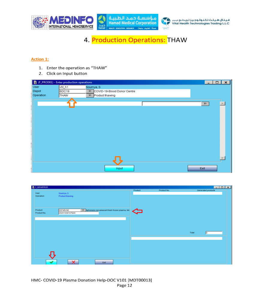
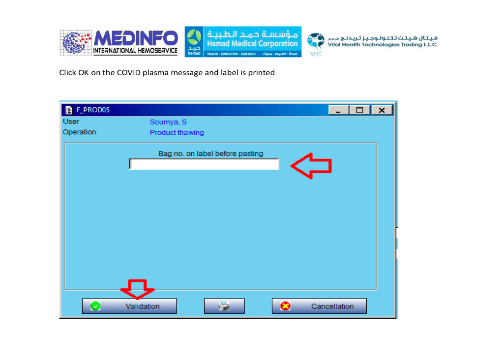
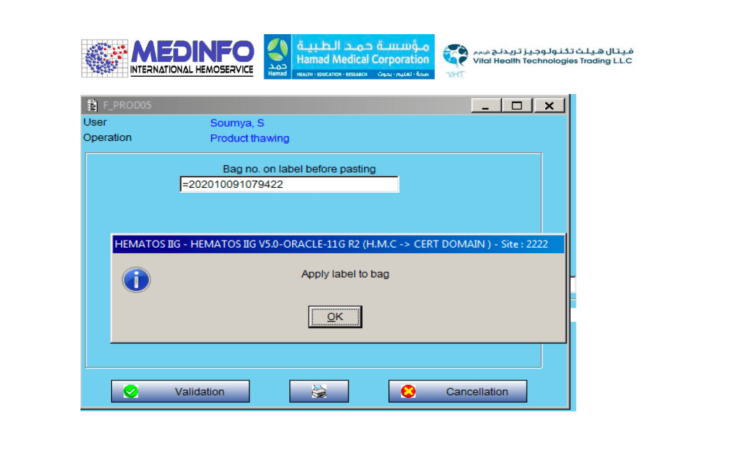
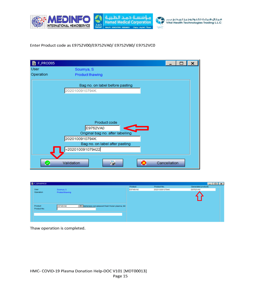
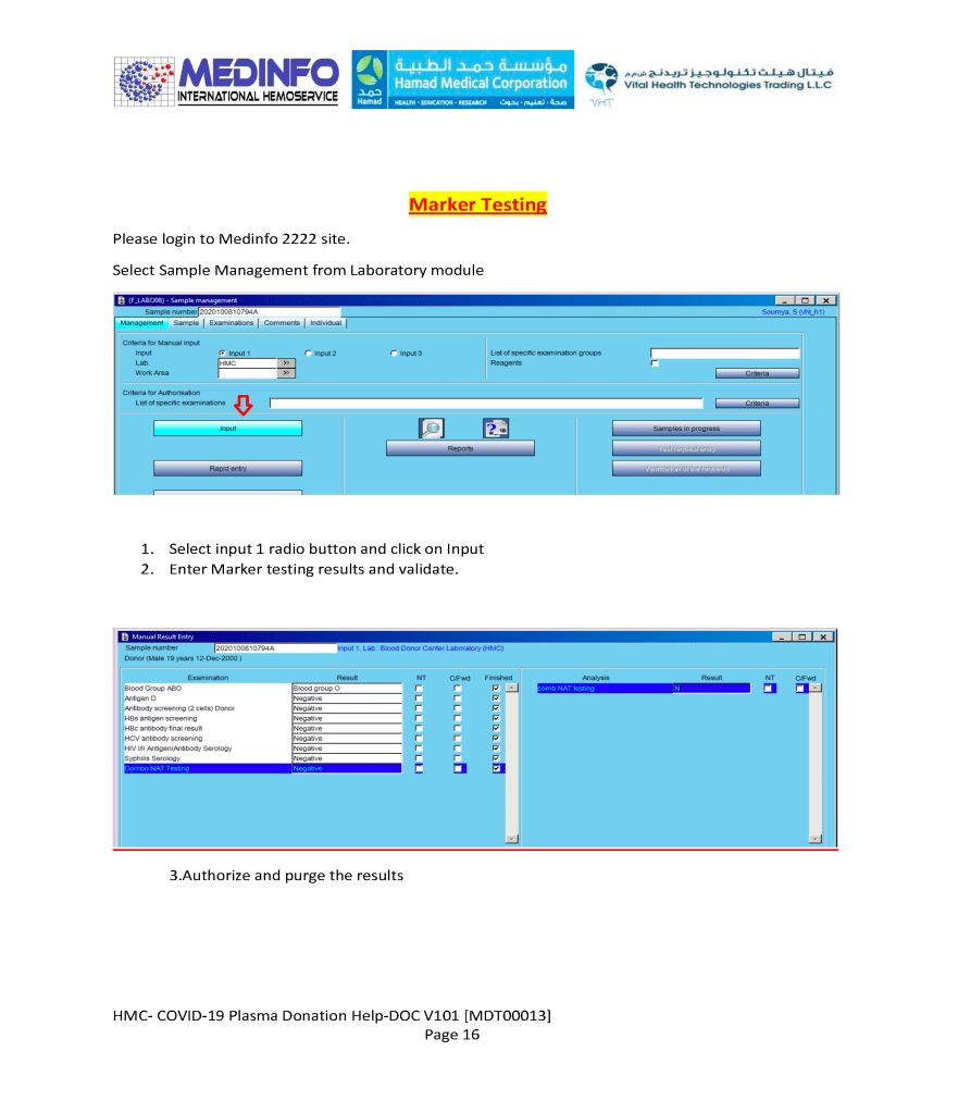
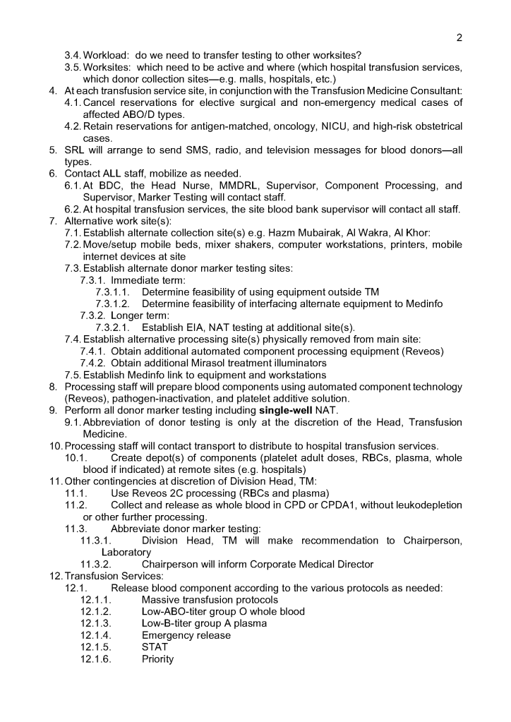
COVID-19 Convalescent Plasma CCP Thawing and Marker Testing

This is a part of a continuing series of posts on the actual Medinfo design of the CCP donation and release processes and covers CCP plasma thawing/labelling and donor marker testing.  It highlights specific changes made for the parallel CCP system.

Thus, the machine interfaces for testing are the same as for regular testing and are not included in this document. Likewise, donor immunohematology testing is the same as for regular donors and is not addressed here

Bedside Monitoring of Transfusion

Once the blood component has left the hospital blood bank, it should be directed transported to the patient’s bedside for immediate transfusion.

At the bedside, the transfusionist (usually a nurse or doctor) must verify the information on the blood component labels (both the ISBT and specific reservation one for the patient).  With a bedside device, this includes:

  1. Scan the patient armband for hospital number
  2. Scan ISBT label:
    1. ABO/D type and any other antigen typings
    2. Blood component type (RBCs, platelets, plasma, etc.)
    3. Expiration date/time of the component
  3. Verify Reservation Label (on back of unit)— centrally performed in blood bank computer system based on #1 and #2 above:
    1. Intended recipient name and hospital number
    2. Compatibility status (compatible, least-incompatible, etc.)
    3. Expiration date/time of the compatibility testing/crossmatch
  4. Record Documentation During Transfusion:
    1. Physical inspection of the unit
    2. Time stamp of transfusion start
    3. Vital signs before starting transfusion
    4. Periodic vital signs during transfusion
    5. Time stamp of stopping transfusion
    6. Any adverse effects during the transfusion
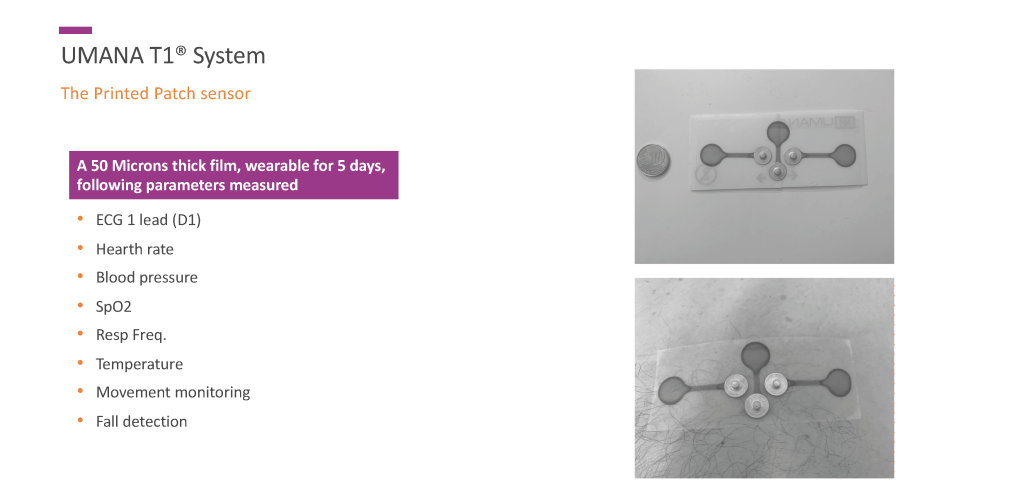
    7. Any adverse effects after the transfusion (remote vital signs, oxygen saturation, EKG lead—if appropriate monitoring device attached to patient (e.g. Umana T1 device).

All of these parameters can be entered into a hand-held device that transmits them to the blood bank computer system (e.g. Medinfo Hemotrace.)

The Blood bank computer system (patient module) should verify that this is the proper unit for the intended recipient and that the transfusion is starting within the reservation limit of the unit and serves to verify the information on the reservation label attached on the back of the blood unit.

With a device such as  the new Umana T1 device from the GPI group, the vital signs together with an EKG lead and oxygen saturation can be automatically uploaded into the handheld device that feeds into the blood bank computer system such as Medinfo Hematos IIG and/or directly transmitted to the blood bank software.  This device can continuously record this information for several days and detect post-transfusion adverse effects such as TRALI/TACO and delayed hemolytic transfusion reactions.

Note:

Some other nursing hand-held devices are available for recording patient data but many cannot read the ISBT unit number or descriptor information.  They also do not check with the blood bank computer system to check if the right unit of blood is being offered to the patient during the validity of the transfusion period (four hours after leaving the blood bank).

Washed RBCs

Note:  This is an updated version of a previous post.

Principle:

Washing RBCs removes plasma and reduces the leukocyte count only by 1 log.  For leukodepletion, we must rely on filtration to reduce the WBCs to less than 1 x 10⁶ per unit according to CE rules.  Red cells or platelets in additive solution contain only minimal plasma (about 35 ml).  There are few definite indications for washing RBCs and it should be rarely necessary.

Policy:

Washing RBCs should only be done in the following circumstances:

  1. Deglycerolization of frozen RBCs.
  2. Severe allergic or anaphylactic reactions to plasma proteins
  3. IgA deficiency with anti-IgA
  4. Paroxysmal nocturnal hemoglobinuria PNH—relative indication (often these patients receive RBCs before the diagnosis of PNH is confirmed)
  5. Transfusing a previously irradiated RBC unit for pediatric use if more than 24 hours has passed since it was irradiated.
  6. Any other time when so designated by a transfusion medicine consultant.

Note:

  1. If anyone requests washed RBCs and it does not fit into one of the above categories, contact the transfusion medicine consultant.
  2. Washed RBCs are NO substitute for leukodepleting RBCs by filtration NOR can they be used in place of irradiation for prophylaxis against transfusion-associated-graft-versus-host disease TAGVHD.  Using the Reveos automated component processing system, all components are leukodepleted—RBCs are released in SAGM.

Reference:

Standards for Blood Banks and Transfusion Services, Current Edition, AABB, Bethesda, MD, USA

Interpretation of Donor Ortho Blood D Blood Group Reagents

Principle:

For the purpose of typing blood donors, we want to detect weak and partial D types and consider them as D-positive since even a portion of the D molecule is immunogenic and sensitization to it may cause anti-D hemolytic disease of the newborn.

Background:

Ortho Diagnostics Reagents use three different monoclonal antibody cocktails that react variably with the antigen D (Rh1)—these are found on TWO (2) cards:  Anti-A/B/A,B/D/CDE and Anti-DVI:

Anti-D/Anti-RH1—IgM monoclonal antibody clone D7B8 can detect most examples of weak and partial D including weak D types 1, 2, 3, 4.0, and D categories II, III, IV, V, VII, DBT, and R0Har.  It does NOT detect category VI.  Retest positive reactions of 2+ or less by an alternate method.  It may show different serologic activity compared to other D typing reagents.

Anti-CDE/Anti-RH1,2,3—IgM monoclonal blend of clone MS24 (anti-C), clone MAD2 (Anti-D), and clone C2 (Anti-E) can detect most cells expressing C, D, or E antigens.  Most examples of partial D including DVI and weak D express C or E antigens and will be detected directly by the included anti-D or indirectly by the anti-C or anti-E in the cocktail.  It does NOT detect Rh:33 (R0Har).

Anti-DVI/Anti-RH1 will agglutinate cells with a DVI phenotype, analogous to our previous DVI+ reagents.

Policy:

  1. Follow the manufacturer’s instruction for storage, handling, and usage of all reagents.
  2. If the D-control is positive, the reactions are indeterminate, repeat by another method.
  3. For donors, run all three anti-D reagents listed—do not use the patient typing algorithm or reagents.
  4. Use the following table for interpretation and further actions if needed:
Pattern #Anti-D/D7B8Anti-CDEAnti-DVID-Interpretation
1PositivePositivePositiveD-positive
2PositivePositiveNegativeDo additional testing
3PositiveNegativeNegativeDo additional testing
4NegativeNegativePositiveDo additional testing
5PositiveNegativePositiveDo additional testing
6NegativePositivePositiveD-positive, probable DVI variant
7NegativePositiveNegativeD-negative, probable rare genotypes r’ and/or r’’
8NegativeNegativeNegativeD-Negative

If the reaction is 2+ or less with the Ortho anti-D/D7B8 reagent or 1+ with either the CDE or DVI reagents is patterns 2, 3, 4, or 5 above, repeat by another manufacturer’s reagents, including DVI+ and DVI- sensitivities.

Medinfo-Ortho interface settings for Blood Donor Center:

Anti-D/D7B8Anti-CDED-Interpretation
3,41,2,3,4D-positive
3,40Indeterminate
00Indeterminate
01,2,3,4Do DVI REFLEX
REFLEX DVI If DVI-pos, then D-positive
REFLEX DVI If DVI-neg, then D-negative
00D-negative
~~Indeterminate

~ means any other result

Note all of the following:

  1. No reagents may be able to detect all D variants.
  2. ~ means any other value for that reagent (e.g. anti-D/D7B8 reactions 1, 2, mf, hemolyzed)
  3. Note that this new algorithm makes a 2+ reactivity with Anti-D/D7B8 as indeterminate.

References:

  1. Product Insert, Anti-A/B/A,B/D/CDE/Control Card, Revised January, 2015, Ortho Clinical Diagnostics, High Wycombe, Buckinghamshire/UK
  2. Publication J55650_EN, Instructions for Use, Blood Grouping Reagent Ortho Sera Anti-D(DVI) (Anti-RH1), Version 2.0, 2015-07-30, Alba Bioscience, Edinburgh, UK
  3. Standards for Blood Banks and Transfusion Services, 29th Edition, AABB, Bethesda, MD, USA

Opinion: Ready after Fellowship?

I was recently interviewing a candidate for consultant in Transfusion Medicine.  Several months previously he had completed a fellowship in Transfusion Medicine in the United States.  He was applying for a position in my hospital in Qatar, which included seven hospitals and a blood donor center.  He had no training in donor management or therapeutic apheresis.

The successful candidate was to rotate on-call to cover all hospitals and the blood donor center.  He had never worked outside the United States.  Routinely, he did not review antibody panels since those workups were usually sent to the local blood provider there.  In his training, he had strictly followed US FDA and American version of AABB Standards.  His training center did not routinely do extended phenotypes (C, c, E, e, and Kell).  Extra testing and phenotyping had to be explicitly ordered by the clinician to get reimbursement.  Thus, there was no prophylactic antigen matching done on patients.  He did not feel comfortable reviewing antibody panels.

He had no experience with universal leukodepletion, pathogen-inactivation, platelet additive solutions, or automated component production such as the Terumo BCT Reveos.  He did not interpret donor marker testing results.

On the contrary in our organization, the transfusion medicine physician had to review all antibody panels (usually he was the most knowledgeable person for this).  We followed the Council of Europe CE and other practices that did prophylactic antigen matching.  We were also in charge of donor qualification and therapeutic apheresis and reviewed any product deviations from the Reveos and donor marker testing.

Clearly, this candidate did not practice transfusion medicine in the way that was necessary for our operations.  We could not cut him loose and make him responsible for a hospital transfusion service or the blood donor center.

Let us contrast this candidate for one being recruited for anatomic pathology/histopathology.  Grossing specimens, performing frozen sections, reading slides, diagnosing cases are the same everywhere in the world.  After completing his American certification, he could perform his profession almost anywhere in the world.

Transfusion medicine practices need to be localized and the selection of blood components and donor qualification are different.  Most of the world does not follow US FDA and has access to blood components, tests, and other technology that is different and maybe more advanced than his training in the USA.

I gave him a clinical scenario to interpret.  An AB patient with anti-K needs to be transfused with plasma.  Are there any special requirements for the plasma?  What if the only AB donor had anti-K would you use it?  What if the only RBCs available had not been phenotyped for Kell?  What would you do?

He did not know that we discard plasma with clinically significant alloantibodies routinely.  He did not want to phenotype the RBC unit for this patient since this had not been explicitly ordered by the clinician.

My recommendation was not to hire this candidate if there were others who had worked in European or similar systems to our own practices.  In effect, to use this physician, he would have to undergo a mini-fellowship to learn our practices since they were contrary to ours.  Unfortunately, we were very short-staffed and did not have resources to offer this training.

In summary, blood bank practices are very localized.  If you are considering to hire staff from other countries not following your standards, you must assess if the candidate is flexible to change his practices and/or whether you have the resources to train the physician.