Inter-Depot Transfer, Blood Delivery, Type and Antigen Matching

This is an update of a previous post.

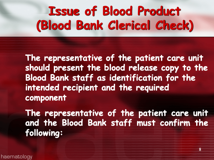
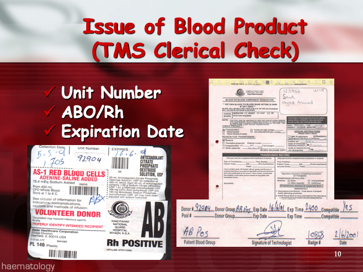
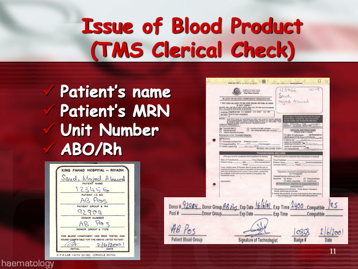
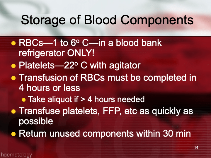
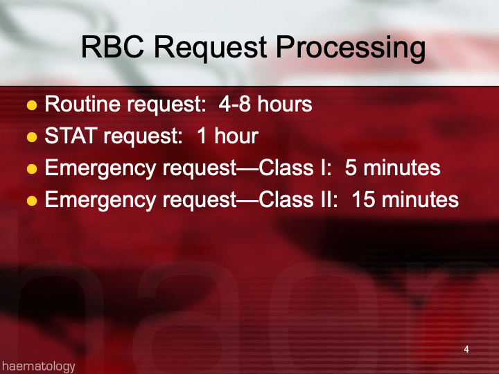
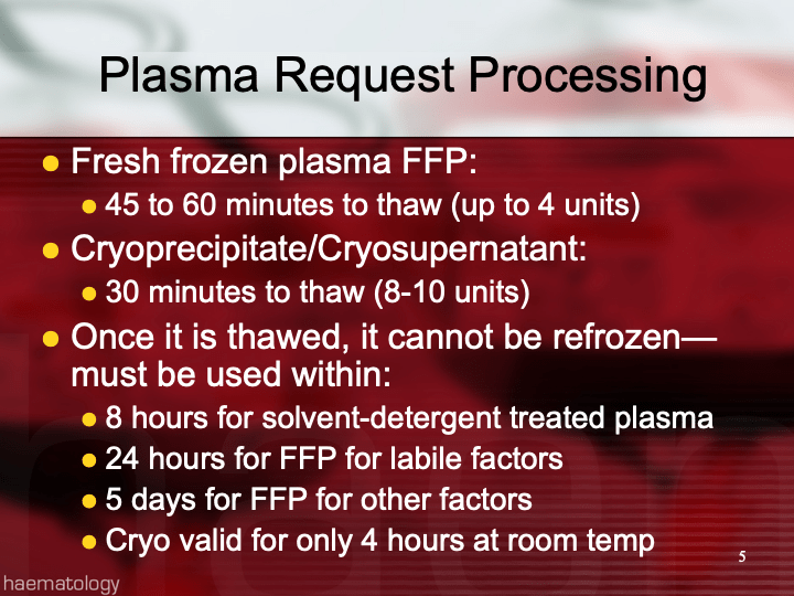
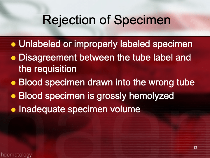
The final components from the component preparation center may be sent to various depots (freestanding location and/or hospital blood banks.  There should be complete traceability for every step (from donor reception, collection, testing, and processing) transport between locations, and finally the exact storage site, which might include which refrigerator/freezer/incubator and even shelf/position number for each component is stored.  The end of that document showed rules for type/antigen matching.

For disaster planning, rapid inventory enumeration by type is very important.  This can be very time-consuming manually.  With our Medinfo Hematos blood bank system, we could quickly get total inventory across the Qatar or by hospital in less than one minute.  We could also quickly find antigen-matched units across the system and reserve it at any one site for another if necessary.

Smart blood bank dispensing refrigerators, as offered by Haemonetics and Angelatoni, may also serve as depots and take the place of a hospital blood bank for some dispensing.  These solutions can also capture vital information about the storage conditions of the components and prevent release if the storage criteria are not met.  They can also interface with blood bank computer systems and use the main system’s logic for the dispensation rules.

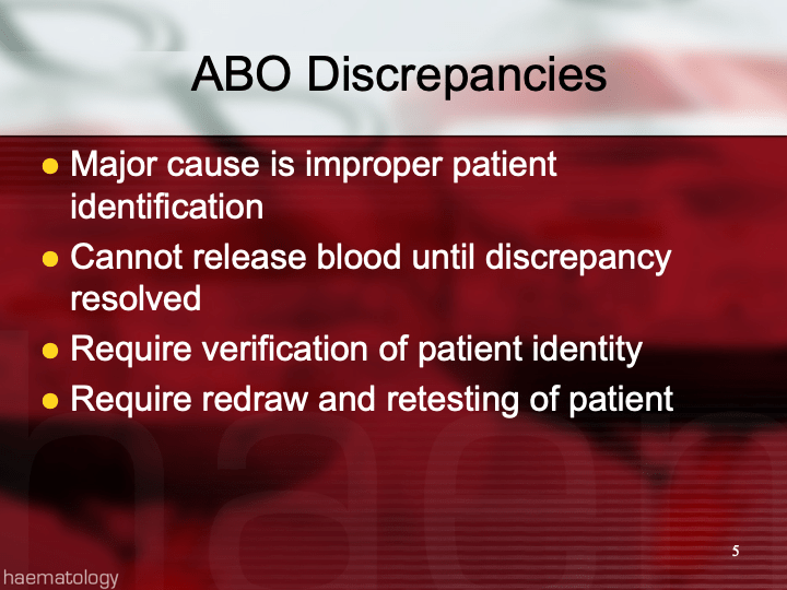
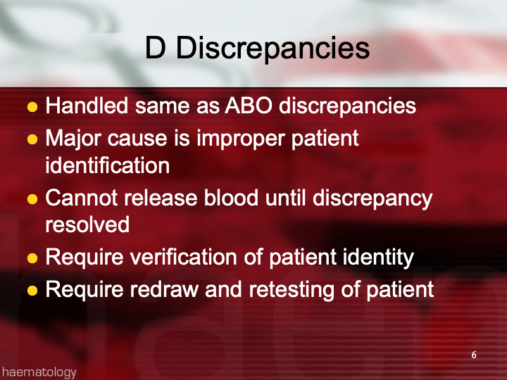
Upon receipt at the hospitals from the blood processing center, the forward ABO and D typing must be confirmed.  We used D reagents which detected partial D so we would call such donor units as D-positive.  However, if a patient type reagent insensitive to partial D types is used, it is possible for a unit to be typed as D-negative whereas in the donor center it might be D-positive.  Sometimes, nothing types consistently as D-positive:  all you can say is that with a particular reagent and lot number, there is or isn’t reactivity.

The greatest complexity is for RBCs since potentially so many antigens exist.  Criteria for matching/ignoring certain antigens must be made.  Critically significant antibodies such as the Kell, Duffy, Kidd, and certain Rh (D and c) must be antigen matched.  A robust blood bank computer system can enforce these rules.

For other components, antigen/typing may be less important.  In fact, in most situations, any type of platelets can be given to anyone (except neonates).  Despite the potentially incompatible plasma, there is rarely significant hemolysis.  In fact, if pooling platelets without regard to blood types is done, a platelet transfusion is a common cause of a positive direct antiglobulin test DAT—something that is not clinically significant.  No one died of a positive DAT by itself for this reason.

Specific rules for compatible plasma types are important, but nowadays, low-anti-B-titer group A plasma may be used like universal AB plasma.  The challenge is to be able to perform the ABO titration (specifically anti-B) quickly—titration can be a slow process, even with automated equipment.  A similar situation for low-titer, universal group O whole blood requires both anti-A and anti-B titration (I will return to this topic in a future post).

Post RhIG Use Without Antibody Identification

Rh immunoprophylaxis has big effect on the Transfusion Medicine operations.  With a very active obstetrics program, there are numerous prenatal blood bank workups and administrations of Rh immune globulin RhIG—and consequently many antibody identifications.  In fact at my previous position, the largest single source of antibody workups was from patients post-RhIG administration.

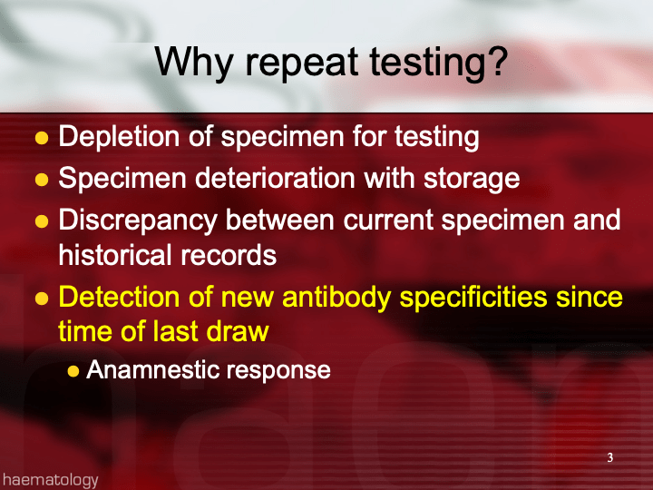
As a result of the antenatal RhIG use, there were numerous positive antibody screens.  Our policy had been to always do a full antibody identification, both AHG and enzyme panels, and this made up the bulk of our antibody testing.  In my experience, I have found other antibody specificities in some of these patients, including anti-Kell and anti-c, both of which could be very clinically significant.

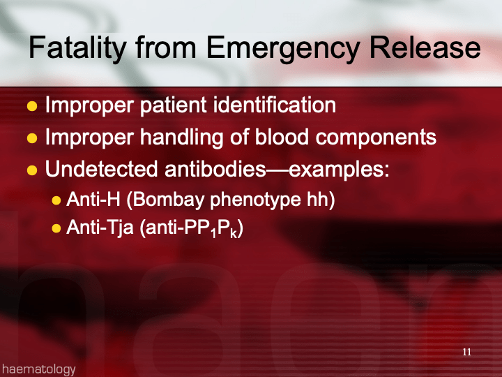
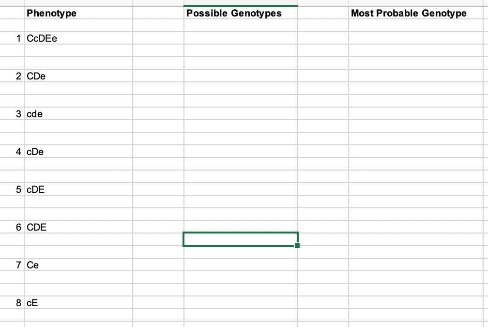
As part of the workup, we also did ABO and extended Rh and Kell phenotyping, and thus identified rare Rh phenotypes (r’r’, -D-, etc.) and several Bombay phenotypes.

Unfortunately, I have been at institutions where they assumed a positive antibody screen in a patient with recent RhIG administration was passive anti-D and did NOT perform further testing.

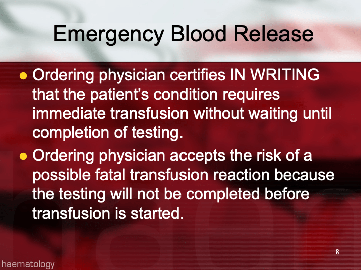
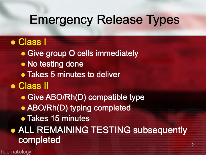
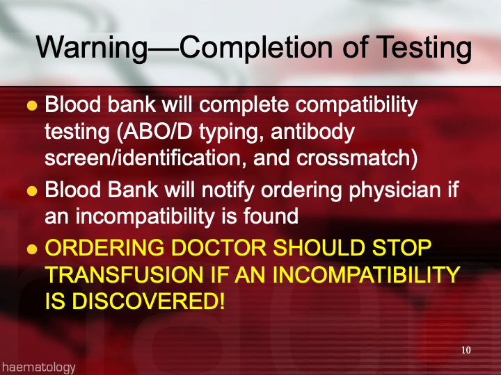
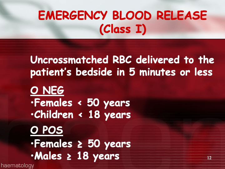
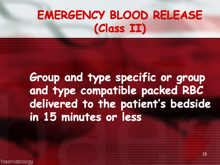
If the patient needs blood and the antibody screen is positive, the antibody workup must be performed for routine release.  This means about a 20-30 minutes delay in release until the antibody panels are completed and reviewed.  Otherwise, blood can be released through an emergency protocol if the clinician accepts responsibility for incomplete testing at the time of release.

Many clinicians refused to do this emergency release and insisted on waiting for the antibody identification before accepting RBC components.  They would not take the responsibility for the emergency release.

If we had done the antibody workup in these RhIG patients, we would have identified the anti-D, and I as the transfusion medicine physician would then advise the clinician to accept emergency release of RBCs while we complete the antibody workup.

For this reason alone, I insisted on a full antibody workup for all RhIG patients RhIG who exhibit a positive antibody screen.  I would be comfortable in recommending emergency release if I had results of the previous antibody identification showing passive anti-D.  The delay in release could adversely affect the patient’s outcome if there is active bleeding.

In summary, if a patient has a positive antibody screen post RhIG administration, you should still do ABID to rule out other antibodies and facilitate release of RBCs to a bleeding patient.

Process: Donor Collection

Process:  Donor Collection

Zeyd Merenkov, MD, FCAP, FASCP

Independent Consultant in Transfusion Medicine

5.4.1 PROCESS DONOR COLLECTION:

Process:

  1. Donors must pass and complete all previous processes in the donor workflow (registration, questionnaire, and physical examination) before the collection process begins.
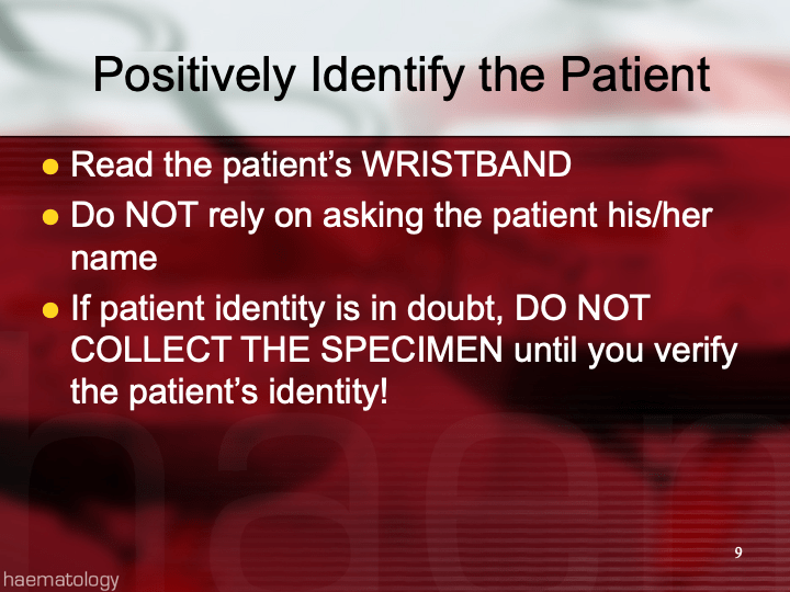
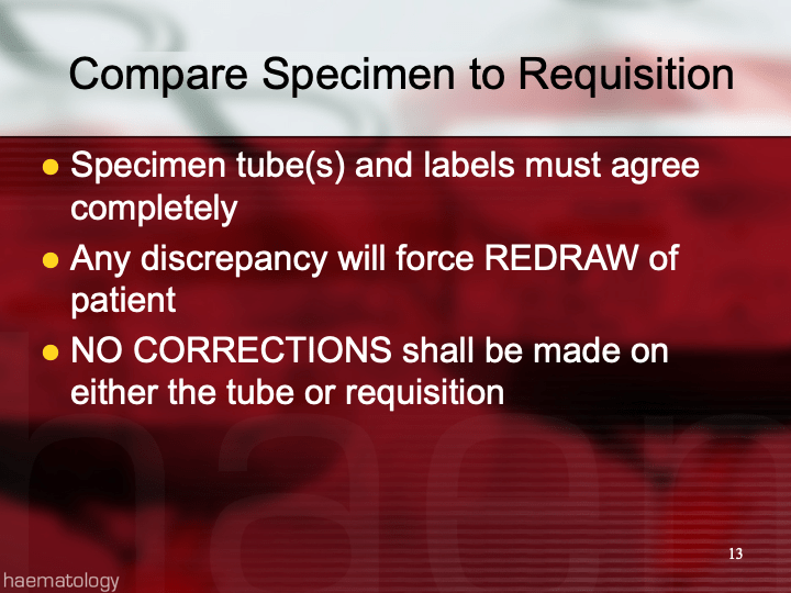
  2. The donor is positively identified by a designated picture ID and Hematos donor consent form with specimen/encounter number and barcode.
  3. Donor staff checks and prepares a suitable vein
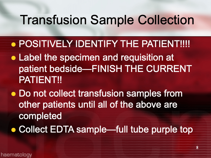
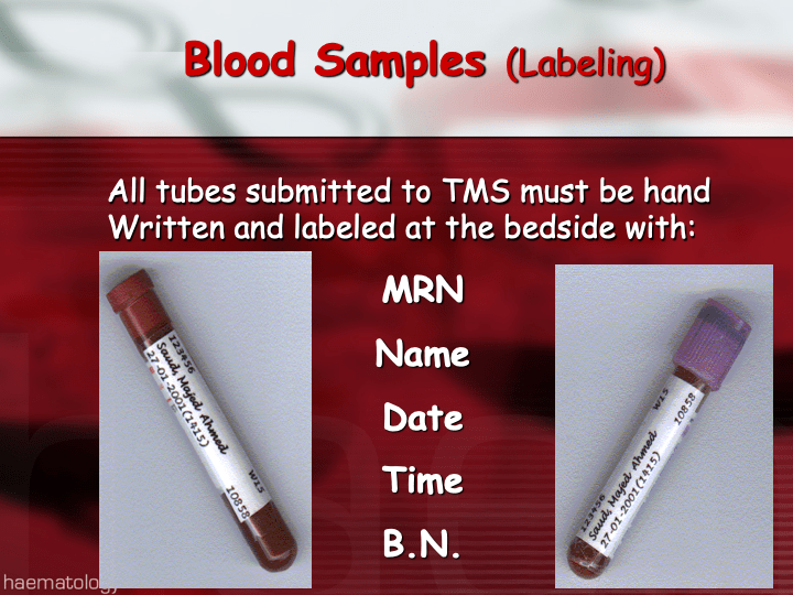
  4. Donor staff collects/labels specimens and the whole blood or apheresis components AT THE DONOR’S BEDSIDE.
  5. Donor reactions are assessed and treated as they occur.
  6. Donors are observed in a post-donation area and given post-donation instructions before discharge.
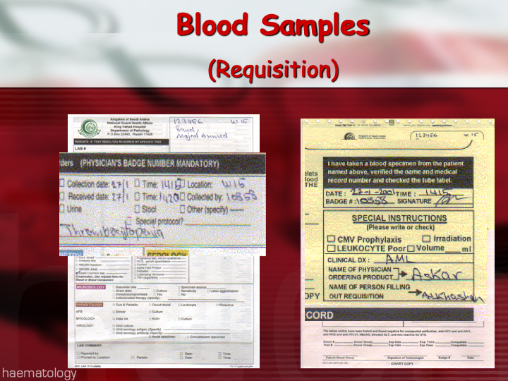
  7. All processes are documented in Hematos IIG.
  8. Donor units and specimens are sent to component processing and donor marker testing.
  9. The collection workstation and equipment are cleaned before starting a new donor collection.

References:

  1. HMC 1001 Setting Specification, Version 1.5, Hematos IIG, Medinfo
  2. Standards for Blood Banks and Transfusion Services, Current Edition, AABB, Bethesda, Maryland, USA

Policy: Donor Collection

5.4 POLICY: DONOR COLLECTION

Policy:

  1. All donors will be positively identified with a picture ID and by their Hematos identifiers (donor ID and session registration/specimen number).
  2. All previous processes (registration, donor deferral database check, questionnaire, and physical examination) must pass.
  3. The donor’s arm veins will be inspected for a suitable donation site and prepared by a suitable aseptic technique.
  4. Whole Blood:
    1. Only whole blood units collected within the specified time interval may be used for component processing.
  5. Apheresis:
    1. Apheresis units will be collected at frequencies to keep the total RBC loss below 200 in any 8-week period.
    2. Only apheresis units collected within the specified time interval may be used for component processing.
  6. Donors will be treated for adverse reactions as needed.
  7. All specimens and donor units will be labeled at the donor’s bedside before starting a new donor collection.
  8. Donors will be monitored post-donation for a reasonable interval before discharge.
  9. All processes will be documented in the Hematos blood bank computer system.
  10. All equipment and supplies will be used according to manufacturer’s instructions.
  11. The collection workstation and equipment will be cleaned before starting the next donor.
  12. All policies, processes, and procedures must comply with Qatari, HMC, and applicable accreditation standards (i.e. AABB, CAP, and JCI).

References:

  1. HMC 1001 Setting Specification, Version 1.5, Hematos IIG, Medinfo
  2. Standards for Blood Banks and Transfusion Services, Current Edition, AABB, Bethesda, Maryland, USA, October 2013