Time: Appreciating How Long Testing Takes

I have had many medical students, residents, and fellows rotate through my Transfusion Medicine department.  Hardly anyone has had any interest in making my discipline his/her career.  It is a required rotation or an “easy” rotation during which the trainee may take his vacation.  The trainee will cram for the examination and then promptly forget it.

I left practice in the USA in 1990, in what I consider the golden age of laboratory medicine.  We had supervisors for each laboratory section.  In the blood bank, we had many staff with SBBs or who were SBB students.  We were very self-sufficient in handling immunohematology problems except for rare blood types or antibodies to high incidence/prevalence antigens.

When I returned to visit my old laboratory.  I sensed a deprofessionalization of the laboratory and blood bank in particular.  Blood Bank now is a cost center, not an area of revenue.  Why hire experienced blood bankers for most hospitals?  Send the antibody workups to the Blood Center.  There are limited jobs for transfusion medicine consultants.  Minimize testing, don’t do extended antigen typings, etc.

Nowadays, I feel like one of the dinosaurs marching into oblivion as in Walt Disney’s Fantasia film, the section called The Rite of Spring.  Who will replace those of us retiring?  Have you ever noted the average age of attendees at the AABB annual convention?  I feel young when I go there (and don’t worry about the gray hair!)

I want to attract new doctors and scientists to Transfusion Medicine.  I really try, but most have no interest and look on their rotations as a necessary evil.

I have lowered my expectations for most medical trainees in Transfusion Medicine.  They don’t like it, they just want to pass it, and move on.  What must I impress them with for their future careers?  What is essential for them to remember?

I have had both pathology and non-pathology trainees.  Surgical and ob/gyn doctors used to spend one month whereas the hematology and pathology residents/fellows spent on average three months.  The few interested in the field might do multiple rotations.

I still gave them lectures on a variety of topics, especially how to transfuse blood components, basic ABO/Rh antigens, compatibility testing, and direct antiglobulin testing.  They would forget most of this, but I wanted them to remember TURN-AROUND-TIMES:

How long does it take to perform the test?

Find compatible blood?

Thaw the plasma?

Release a massive transfusion protocol shipment?

Complete a transfusion reaction workup before releasing more blood?

I am not discouraging people from entering the field, but I am a realist to know that few will share my passion for serology or want to take call on difficult immunohematology cases.  At least if they understand the pressure the technical staff are in and these turn-around-times this will make both their work as clinicians and mine as transfusion medicine more congenial.

Daratumumab Anti-CD38 Interference with Compatibility Testing

Principle:

Daratumumab is a monoclonal antibody that binds to CD38 antigen, which is expressed weakly on the surface of all RBCs.  It may thus cause a positive direct antiglobulin test DAT and so interfere with compatibility testing if an antiglobulin phase is required.

This effect may persist up to 6 months after discontinuing the drug.  The monoclonal antibody does not interfere with routine ABO/D typing.

Special techniques (neutralization of CD38 antibodies by CD38 anti-idiotypic antibodies, or soluble CD38 antigen) may remove the panreactivity but are not generally available.  DTT, a sulfhydryl reagent may denature the native CD38 antigen on RBCs but it should be used under a biologic hood.

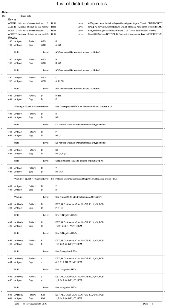
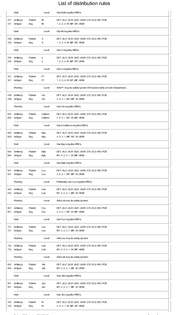
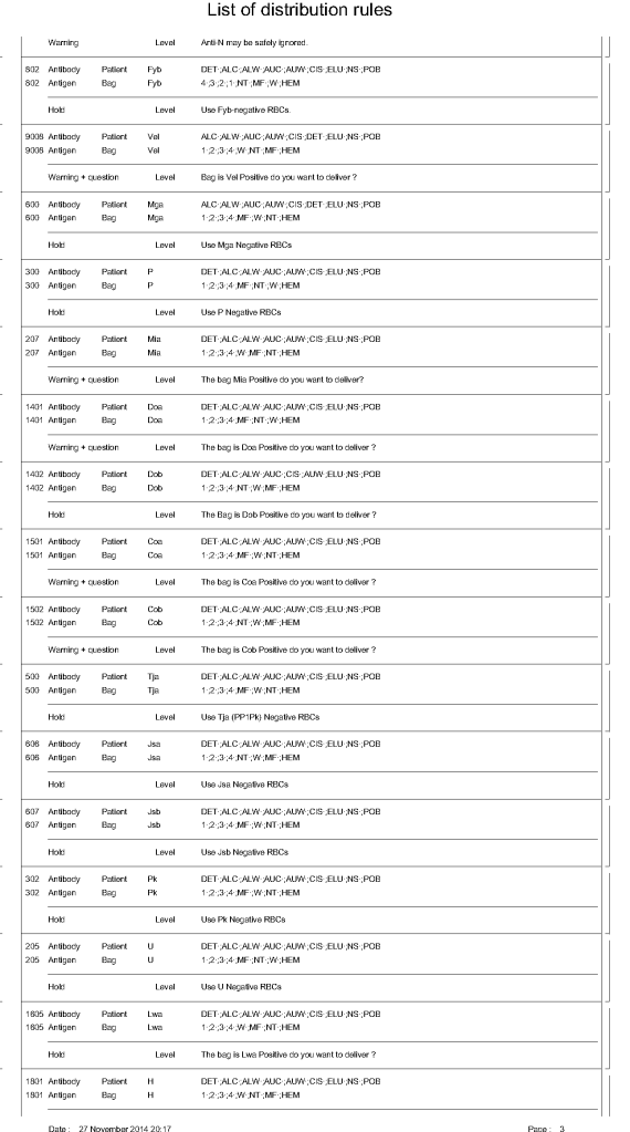
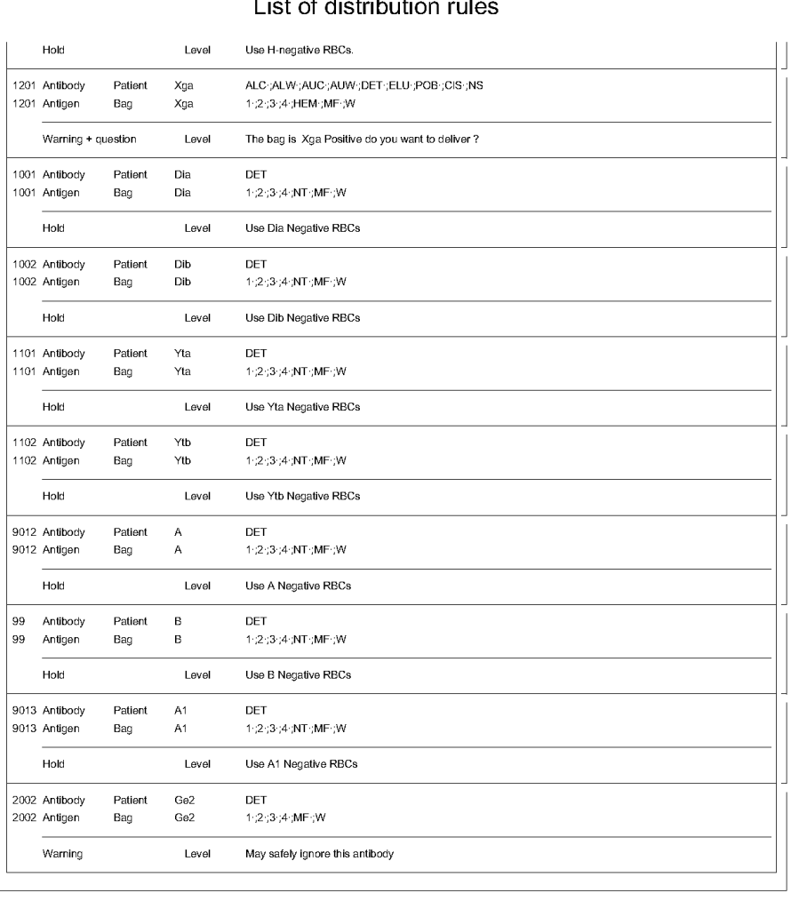
Kell antigens will be denatured so Kell antibodies cannot be detected after treatment so Kell-negative RBCs should be used.  In the Gulf Area, this is about 72% of RBCs. In the Medinfo software a rule to require K-negative RBCs has been built.

Policy:

  1. The clinical services must inform Transfusion Medicine of patients who will be receiving daratumumab therapy BEFORE treatment is started.
  2. Transfusion Medicine staff will enter a general comment (i.e. not associated with a particular result) in the patients Medinfo HIIG record:  PATIENT ON DARATUMUMAB.
  3. If not already done, Transfusion Medicine staff will perform an extended antigen typing:  at least C, c, E, e, K, k, Kpa, Jka, Jkb, Fya, Fyb ,M, N, S, s, Lea, Leb, P1—even if no antibodies are currently identified.
  4. Transfusion Medicine staff will send each such patient’s record to a Transfusion Medicine Physician to determine the blood type including extended antigens to match for future transfusions.
  5. When compatibility testing is requested, perform it as per our SOPs.
  6. If available, prepare DTT-treated cells for testing but realize that this will denature Kell antigens.  Use K-nell RBCs.
    1. Medinfo has a rule to automatically require K-negative RBCs if this medication is used.
  7. Release least “incompatible” RBCs must be approved by the Transfusion Medicine Physician.
  8. When the DAT becomes negative (i.e. up to SIX months after cessation of Daratumumab therapy), routine compatibility testing and RBC selection will apply.

References:

Trick or Treatment, Anti-CD38 Reactivity and How to Treat It, AABB Satellite Symposium transcript, U. Cincinnati and RedMedEd, October, 2015 (attachment)

Processes and Software Building 24: Data Entry Verification

Principle:

This policy outlines steps taken to minimize the risk of data entry errors and is based on a dualistic approach:  review of results by a senior technologist and/or supervisor and various computer safeguards built into the Medinfo Hematos IIG blood bank computer HIIG system.  This policy also discusses the verification (here called authorization) and purge processes of HIIG.

Policy:

  1. Review by senior technical, supervisory, or transfusion medical staff:
    1. Designated test procedures require review by a second technologist before authorization.
    2. Complex immunohematology testing and specimens showing aberrant results (e.g. ABO/D discrepancies) are reviewed by the supervisors or designates and ultimately a transfusion medicine physician before authorization.
  2. Computer system HIIG rules:
    1. Privileges:
      1. System restricts which staff can perform specific tests
    2. Patient/donor identity:
      1. System asks end-users to verify patient/donor identity before starting any access to the patient/donor record.
      2. System performs historical database checking and flags any inconsistencies (e.g. historical ABO/D typing differences, etc.)
    3. Testing:
      1. Only selected staff have privileges to authorize or purge.
      2. ABO/D testing algorithms require entry of reactions, not interpretation of results and are compared to a truth table.
        1. Aberrant results require special review before ABO/D typing results can be authorized/purged.
        2. D-controls must be negative to allow D typing results to be authorized for liquid D-typing reagents.
      3. DAT results require appropriate controls to meet truth-table criteria.
      4. Eluates require last wash to be negative before authorization
    4. Blood components:
      1. Selection of RBC or plasma units requires two independent sample determinations within 72 hours of each other.
      2. ABO-incompatible RBC or FFP/FP24 transfusions are not allowed.
      3. Donors with any detectable antibodies are permanently deferred.
      4. Depending on the patient’s antibody history, release of RBC units may require antigen-matched units.  Examples:
        1. Mandatory matching (only antigen negative matched units allowed—no antigen positive or antigen-untyped units):  Antibodies against H, D, c, K, k, Kpa, Kpb, Jsa, Jsb, Jka, Jkb antigens, anti-PP1Pk
        2. Priority matching (incompatible or untested can be approved by a transfusion medicine physician):  C,E, e, Fya, Fyb, M, S, s
        3. Antigen matching not required:  Lea, Leb, N
      5. Least-incompatible crossmatch require special authorization to release
      6. Protocols to force irradiation or other modified components can be setup in HIIG.
    5. Donors:
      1. Donor tests have same criteria as the same test used in patient testing for controls, etc.
      2. Donor demographics are read directly from the Ministry of Interior database—no manual entry (bar code only used).

References:

  1. Workflows for Hematos IIG (1001 through 1005), 2013
  2. Standards for Blood Banks and Transfusion Services, Current Edition, AABB, Bethesda, MD, USA
  3. Guidelines to the Preparation, Use, and Quality Assurance of Blood Components, European Committee (Partial Agreement) on Blood Transfusion (CD-P-TS), Current Edition

Case Report: Blocking Anti-B

Recently, I had a case where the blocking antibody was not an anti-D, but rather an anti-B in a case of ABO hemolytic disease of the fetus/newborn HDFN.  The D typing result was weak but the D control in the gel was positive so the result was indeterminate.  DAT was 3+ IgG and anti-B was identified in the eluate (mother was antibody screen negative, regular elution panels (group O cells) were negative.  Some of the actual workup follows:

29/7/20

Blocking Antibodies

If there is strong antibody binding to an RBC, this may interfere with a typing reagent attaching to the cell and cause a false-negative, i.e. a “blocking” antibody.  Such cells may interfere with the indirect antiglobulin test IAT, i.e. the antibody screen.  The autocontrol and direct antiglobulin test DAT will be strongly positive.

The manufacturer’s instructions should be strictly followed for using its reagents in the presence of a strongly positive DAT.  If there is no reaction with the typing reagent, the result must be indeterminate.

One could try a (relatively) nondestructive elution method such as gentle-heat elution to remove some of the antibody and then retype the cells.  I have found this to be a simple and effective method for my staff to use.  Just remember that despite being “gentle,” there will still be some hemolysis present, but here it is the cells we are trying to save.

Usually, we find this situation in a neonate born of a mother with anti-D.  The baby has a strong DAT but the D typing is negative.  Check the D control carefully:  if it is positive, the result is indeterminate, try another method.  Usually gel/glass bead methods are subject to less interference.  Finally, there is always the classic saline anti-D!

In Medinfo software with a blocking antibody, a nonnegative control will trigger a manual review of the results. There will be no automatic release.

Here is my process for handling blocking antibodies, which I set up for HMC Doha:

INTERIM POLICY:  ANTIGEN TYPINGS IN PRESENCE OF STRONGLY POSITIVE DIRECT ANTIGLOBULIN TEST (DAT):  RULE OUT BLOCKING ANTIBODY

Principle:

Antigen typing of cells with large amounts of coating antibody (i.e. strongly positive DAT 3-4+) may not always be possible because the bound antibody may block available antigen sites.  This policy is to clarify how to recognize and handle such situations.

Policy:

  1. Always follow the manufacturer’s instructions for the use of the typing reagent.
    1. In particular, note whether a control must be run with the test (e.g. D-control, D-diluent, etc.) or if it is included in the gel or glass bead card.
      1. If a control is required, use exactly what the manufacturer recommends.
      2. DO NOT SUBSTITUTE ANYTHING ELSE AS THE CONTROL!!
  2. Interpret the reactions exactly as the manufacturer indicates.
  3. If the test is invalid because of the control or any other reason, report the antigen typing as indeterminate and send for Transfusion Medicine Physician review.
  4. If the DAT is 3-4+ and the antigen typing shows no reaction (apparent negative), send the case to the Transfusion Medicine Physician for review and final interpretation.  DO NOT ENTER THE RESULT AS NEGATIVE UNLESS THE TMP INSTRUCTS YOU TO DO THIS!!
  5. To rule out a blocking antibody, a special elution to gently remove the coating antibody may be needed so that the RBCs can then be typed (not acid glycine technique—rather, gentle heat elution.)  The Transfusion Medicine Physician will decide whether to do this additional testing.

References:

  1. Standards for Blood Banks and Transfusion Services, Current Edition, AABB, Bethesda, MD, USA
  2. Technical Manual, Current Edition, AABB, Bethesda, MD, USA
  3. Guidelines to the Preparation, Use, and Quality Assurance of Blood Components, European Committee (Partial Agreement) on Blood Transfusion (CD-P-TS), Current Edition

Summary of Accomplishments at HMC 2011-2020

I resigned from HMC on 16/4/20.  Here are a set of my major accomplishments during that period. None of my work after this date has any relationship to HMC.

2011

Established automated component production using Atreus technology, plasma and platelet pathogen inactivation (Mirasol)—made HMC component production Good Manufacturing System GMP compliant

Adopted non-PCR-based NAT technology (Grifols/Novartis Tigress) and Qatar becomes world reference site for this

Based on the above, Qatar can now completely process all whole blood into blood components (red cells, platelets, and plasma) in as little as 5 hours from collection!

2011-2020:

Prepared policies and procedures for the hospital blood banks/transfusion services, blood donor center, therapeutic apheresis, and laboratory information systems to bring HMC in compliance with the Council of Europe, international AABB, and other standards.  I customized our own standards for our local needs based on them.

2012-2013

Implemented custom build of the multilingual blood bank computer system (Medinfo) for both patient and donor services, including development of interfaces to all production equipment including Atreus and Mirasol (world’s first) and a direct link to Ministry of the Interior to obtain patient demographics in English and Arabic—Qatar became the world’s first site to combine fully-interfaced, automated component production with pathogen inactivation:  Qatar becomes world reference site for this.

2013-2014

Built, validated, and implemented laboratory build of hospital information system, Cerner Millennium

2015

Replaced and updated Atreus with Reveos automated component production to allow faster throughput and capacity with a full bidirectional interface (world’s first), introduced platelet-additive solution PAS with pathogen inactivation (Mirasol)—Medinfo interfaces updated to Reveos for all equipment:  this doubles the capacity to process whole blood into components using the same physical space

2015-2019

Updated dedicated blood bank software Medinfo Hematos IIG by several versions using Division Head, LIS, and internally trained Super Users—at great cost savings to HMC by not using outside consultants (e.g. Dell Consulting)

2019

Established column absorption technology using Terumo Optia therapeutic apheresis machine for treatment of ABO-incompatible renal transplants:  I validated using the Ortho Vision MAX to perform ABO antibody titers for this system and correlated it with the reference method at Karolinska Institutet in Stockholm (manual gel) to bring rapid throughput and labor savings—Qatar being the first-site in the world to do this.  We saved money by using the same apheresis machine to use this column absorption technology (no need for second machine to use the columns)

2020

Expedited setup (two weeks total) of COVID-19 convalescent plasma production, initially manual and then fully integrated into the Medinfo computer system as a customized module with separate quarantine collection, production, and transfusion service functions

Other:

I was awarded two HMC Star of Excellence Awards:

2013—Liver Transplantation Transfusion Support

2019—ABO-Incompatible Renal Transplantation Support

TRALI/TACO Policy and Process

The following was my process at HMC Doha for TRALI/TACO.  It includes proactive measures to minimize the risk of TACO and the procedure for surveillance and workup of such cases.

In the Medinfo blood bank computer system, we did not prepare plasma or platelets from female donors.  If approved by a transfusion medicine physician, a manual override was made in exceptional cases (e.g. mother donating platelets for her child in neonatal alloimmune thrombocytopenia cases.).  In some other countries, they do HLA antibody testing to allow females to donate platelets.

I emphasize that the diagnosis of TRALI and/or TACO is clinical, but the transfusion medicine physicians must always consider the possibility whenever there is an adverse effect associated with progressive respiratory distress.

Principle:

Since TACO and TRALI are major causes of serious adverse effects from transfusions, this policy outlines actions being pro-actively taken to mitigate the risks in Transfusion Medicine.  TACO and TRALI may be difficult to distinguish so this policy addresses both.

Objectives:

  1. Implement measures to minimize TRALI and TACO
  2. Track cases of transfusion-associated acute lung injury TRALI and TACO
  3. Develop algorithms for suspected cases of TRALI and TACO

Tracking:

  1. All transfusion reactions are reviewed by the Division Head, Transfusion Medicine or his designee on a STAT basis, 24 hours a day, 7 days a week
  2. Any reactions with respiratory distress are reported as “rule-out TRALI/TAC” to the clinician
  3. All transfusion reactions are recorded in Medinfo HIIG for tracking and reporting.

Risk Management:

  1. Female blood donors routinely are only used for making RBC components (i.e. not for FFP, cryoprecipitate, or platelets).
  2. RBCs are in additive solution SAGM so only 35 ml residual plasma is present per unit.
  3. All platelet components are in platelet additive solution with only 35 ml residual plasma per component.
  4. All platelets and plasma are pathogen-inactivated which may reduce the risk of TRALI.
  5. If the female has a rare phenotype (e.g. IgA deficient, rare platelet antigen typing) she will only be considered for a directed donation of platelets or FFP/cryoprecipitate for that special-needs patient if she does not have HLA antibodies (anti-human-neutrophil antibody testing to be implemented in such cases when it is available on-site).
  6. Solvent-detergent treated plasma SDP is available for patients with a confirmed or suspected history of TRALI.
  7. All cellular components are leukodepleted (< 1E6 residual WBCs/component as per CE Standards) in the blood bank at the time of production.
  8. Blood bank computer system Medinfo Hematos IIG limits the number of components released at any one time (excluding emergencies).

Notifications:

  1. Transfusion Medicine TM will notify the outside blood supplier of any units implicated or associated with TRALI.
  2. A transfusion medicine physician will notify the most responsible physician of any workup results suggesting the possibility of TRALI/TACO.
  3. TM will notify all donors of their disqualification from blood donation based on the following algorithm.

Algorithm for Diagnosis and Management of Donors:

  1. Evaluation of the Donor and Recipient in Suspected TRALI:
    1. The medical technologist will process all transfusion reactions as STAT and immediately contact the TMS Director or physician designate with the results.
    2. The medical technologist will convey information to the TMS Director or designate about ALL blood components issued recently, especially in the last 6 hours prior to the event.
    3. The TMS Director or designate will specifically check if there is evidence of respiratory distress listed on the transfusion reaction investigation form.  If so, he will contact the responsible clinician immediately for further assessment.
    4. If the signs and symptoms suggest ALI (see Table 1 above), the TMS Director or designate will inquiry about the left atrial pressure to rule out left-sided heart failure as a cause for the pulmonary edema.
    5. Based on the clinical information, the TMS Director or designate may elect to order any or all of the following tests if available:
      1. Quarantine all remaining components from possibly implicated/associated donor(s) while the workup is in progress.
      2. Recipient HLA, platelet, and/or granulocyte antibody screen, or a crossmatch between recipient plasma/serum and donor leukocytes
      3. Donor HLA and/or platelet antibody screen, granulocyte antibody screen, crossmatch donor plasma/serum and recipient leukocytes, inter-donor crossmatch between plasma/serum of one and leukocytes of another donor.
      4. The usual algorithm to be followed is as follows:
        1. Consult patient medical record and clinical care physician to determine if the diagnosis of TRALI is likely.
        2. Check all components transfused within 6 hours prior to the onset of symptoms.’
        3. Immediately quarantine other components from the same donations and contact outside blood suppliers if indicated.
        4. Obtain donor antibody testing of only highly suspect cases, based on the clinical manifestation and initial diagnostic tests:
          1. If multiple units transfused within hours, only investigate components donated by multiparous females and/or last two units transfused.
          2. First test for presence of HLA class I and class II antibodies in donor components.
          3. If antibody positive, HLA type recipient’s lymphocytes to detect corresponding antigen or perform crossmatch with donor plasma and recipient lymphocytes.
          4. If HLA antibody negative, proceed with neutrophil-specific antibody testing of donor plasma.
        5. If matching antigen-antibody identified or if positive crossmatch, defer implicated donor immediately.
        6. If no such concordance found or if crossmatch is negative, donor eligible to continue donating.
        7. If no antibodies found in donor plasma, test recipient plasma for antibodies to HLA class I and II antigens:
          1. If recipient antibody positive, HLA type donor’s lymphocytes to detect corresponding antigen or perform crossmatch with recipient plasma and donor lymphocytes.
          2. If recipient HLA antibody negative, proceed with neutrophil-specific antibody testing of recipient plasma
  2. Donor Disposition:
    1. For donors implicated in TRALI or associated with multiple events of TRALI, one or more of the following options may be selected at the discretion of the Head, Transfusion Medicine or designate:
      1. Defer donor from donation
      2. Divert plasma for fractionation or discard plasma from future whole blood donations from that Blood and Apheresis Donor Main Questionnaire
      3. Manufacture no platelet or plasma components from that donor
      4. Wash or freeze/deglycerolize RBCs from that donor
      5. Permanently defer the donor from future plasmapheresis or plateletpheresis donations
      6. Evaluate the previous donations from that Blood and Apheresis Donor Main Questionnaire  Avoid giving the same recipient future transfusions from the same donor implicated in TRALI
      7. If the implicated unit(s) are from another facility, that blood center should be notified to initiate a workup for possible TRALI in the donor.
  3. Interpretation:
    1. The diagnosis of TRALI is not clear-cut:
      1. The AABB interim standard does not apply.  It is at the discretion of the TMS Director or designate whether to conduct donor assessments.
    2. The donor is associated with a single event of TRALI:
      1. This applies where the diagnosis of TRALI has been established based on clinical and radiographic findings:
      2. Each donor from each and every component associated with TRALI must be identified and traced.
      3. Co-components from the current donation and components from previous donations should be evaluated for recipient complications.
      4. The donors medical history should be evaluated for previous pregnancies, transfusions or other events that may have resulted in antibody development.
      5. Based on the results of this investigation, the Head, Transfusion Medicine or designate should decide:
        1. Whether to perform laboratory testing
        2. Whether to discard the remaining blood components from the donor
        3. Whether to allow or indefinitely defer the donor
    3. The donor is associated with multiple events of TRALI:
      1. This applies where the diagnosis of TRALI has been established based on clinical and radiographic findings:
      2. Each donor from each and every component associated with TRALI must be identified and traced.
      3. Co-components from the current donation and components from previous donations should be evaluated for recipient complications.
      4. The donors medical history should be evaluated for previous pregnancies, transfusions or other events that may have resulted in antibody development.
      5. Based on the results of this investigation, the TMS Director or designate should decide:
        1. Whether to perform laboratory testing
        2. Whether to discard the remaining blood components from the donor
        3. Whether to allow or indefinitely defer the donor
    4. Triage based on laboratory testing for TRALI:
      1. The donor associated with TRALI is antibody-negative:
        1. The donor may continue to donate.
      2. The donor associated with TRALI is antibody-positive but the specificity is NOT directed against a recipient antigen by either antigen typing or crossmatching (i.e. the donor is NOT implicated in TRALI—see definition above):
        1. Indefinitely defer the donor from all donations OR
        2. Allow donation of washed/frozen-deglycerolized RBCs only
      3. The donor is implicated in TRALI (see definition above):
        1. Indefinitely defer the donor from all donations OR
        2. Allow donation of washed/frozen-deglycerolized RBCs only
      4. The recipient has antibodies implicated in TRALI (determined by crossmatch or antibodies directed against specific HLA class I, HLA class 2, and/or human neutrophil antigens):
        1. The recipient must receive leukodepleted blood components
    5. TACO
      1. TACO is due to cardiac overload.  Our mitigations are to restrict release of the number of components outside emergency events.

References:

  1. AABB Association Bulletin 14-02, TRALI, Bethesda, MD, USA
  2. Han Y. and Goldfinger D., Transfusion Medicine TM 07-5 (TM-297) Checksample, American Society for Clinical Pathology, Chicago, IL, USA. July 2007
  3. Goldman M, Webert, KE, Arnold DM, et al., Transfusion Med Rev  2005; 19:2-31.
  4. Fung YL, Goodison KA, Wong JK, Minchinton RM., Investigating Transfusion-Related Acute Lung Injury (TRALI), Intern Med J. 2003 Jul;33(7):286-90.
  5. Standards for Blood Banks and Transfusion Services, Current Edition, AABB, Bethesda, MD, USA
  6. AABB Association Bulletin #05-09, Transfusion-Associated Acute Lung Injury, 11/8/05
  7. AABB Association Bulletin #05-04, Proposed Interim Standard for Deferral of Donors Implicated in TRALI, 9/3/05.
  8. TRM.42110, CAP Transfusion Medicine Checklist, 15/6/09

Processes and Software Building 22: Review of Laboratory Reporting Formats

Principle:

In accordance with the College of American Pathologists’ accreditation standards, all report structures (content, formatting) are reviewed at least biannually and upon modification by staff designated by the Chairperson, DLMP, here the Division Head, Transfusion Medicine/Laboratory Information System.

Policy:

  1. Responsibilities:
    1. The Chairperson, DLMP is ultimately responsible for the report formats, electronic and paper (if applicable) across all areas/divisions/sections of the department.
    2. The Chairperson, DLMP delegates the responsibility for this review to the Head, Laboratory Information Systems LIS.
    3. The Head, LIS reviews/approves the final reporting formats after review/acceptance of the formats by the Division and Section Heads for their respective areas.
  2. Content of the reports:
    1. Headers and footers as required, especially for paper reports
      1. Headers will include full patient name and a unique alphanumerical identifier, age, location, sex, date of testing/reporting, location, and ordering physician.
      2. Footers will include contact information for the site performing the testing and for the Chairperson, DLMP.
    2. Body of the report will include:
      1. Test results
      2. Flags
      3. Reference range
      4. Order and result comments
      5. Corrected/amended/appended results will be clearly marked, including any changes from the initial reports.
  3. Documentation of Review:
    1. Screenshots (electronic) or printout (paper formats) will be collated for one example of each test and reviewed by the Division/Section Heads and documented with a signature/stamp for each test result.
    2. Upon acceptance by the Division/Section Head, a cover letter summarizing the acceptance will be signed, stamped, and dated.
    3. The completed documentation will then be submitted to the Head, LIS for final review and approval.

Reference:

GEN.41077, Content/Format Report Review, CAP Checklist, Current Edition

The following is a sample report I prepared during my tenure at HMC Doha:

On the left-hand side of this composite PDF there are embedded attachments, which can be accessed by clicking on each one.  In this document,  I have shown a sample page from the actual screenshots generated.  The data showing is a dummy test patient (no real patient data is exposed).

Note that our design of Medinfo did not include printing copies of reports.  The only available reports were the screens.  Staff outside Transfusion Medicine viewed the blood bank reports through a separate database viewer.  In the example below, the Medinfo screen appears first followed by the EMR Viewer report.

Diamed/Biorad Profile Cards 1-2-3 for Extended Antigen Typing

Processes and Software Building—Part 21

In any case with nonspecific antibodies and for all new patients who will require chronic transfusions, I perform extended Rh (CcEe)/Kell and the three Diamed (now Biorad) profile cards.

It is very easy in Medinfo to write a process for any group of antigen typings as long as you know the manufacturer’s criteria for accepting results.  Some cards have controls, others do not.  In the latter case, the “control” is a negative reaction in the card or series of cards of the same type.

In Medinfo, one can also look for errors in using the card:  In Profile Card 3, i.e. the MNSsFyaFyb card, one must reject the card if no reactions in any well appear:  Did the technologist forget to add the cells or reagents?  Did he/she use the wrong diluent (i.e. bromelin enzyme) which would destroy the labile antigens?

One can set the acceptable range of reactivity, flag for mixed field, etc. and record these findings in the official record.  One can define which reactions you will accept an automatic reading of the card.  For the other readings, one can force a manual review and result entry.

Note that Profile Cards 1 and 2 both have an internal control whereas Profile Card 3 (enzyme-labile antigens) does not.

Here are the processes for all three profile cards:

23/7/20

Immediate Spin Crossmatch

Principle:

Immediate-spin crossmatch ISXM is an abbreviated compatibility testing that detects most ABO incompatibility.  AABB Standards restricts its use to specific situations as indicated in the Policy section below.

Policy:

  1. Applicability:
    1. ISXM may only be used when ALL of the following conditions are met:
      1. Negative antibody screen
      1. No history of antibodies
      1. No ABO/D antigen typing discrepancies
      1. Less than two (2) ABO/D determinations are on-file.
    2. Use the computer/electronic crossmatch whenever its specific criteria are met.
  2. Testing:
    1. ISXM consists of reacting an RBC suspension with patient/serum or plasma, centrifuging, and immediately reading for agglutination or hemolysis.  THERE IS NO INCUBATION STEP, NO USE OF ANTIHUMAN GLOBULIN AHG REAGENT.
    2. If any hemolysis or agglutination is detected, the crossmatch is incompatible:
      1. Repeat the ISXM.
      2. Repeat the patient ABO/D AND unit ABO/D typings.
      3. Repeat the antibody screen.
      4. Perform full AHG crossmatch AND AHG/enzyme antibody panels.
      5. Refer the case to senior technical staff or a Transfusion Medicine consultant for further instructions.
        1. Case review will be documented in Medinfo HIIG computer system.
      6. DO NOT RELEASE THE RBCS UNTIL SO INSTRUCTED BY SENIOR STAFF.

References:

  1. Technical Manual, Current Edition, AABB, Bethesda, MD, USA
  2. Standards for Blood Banks and Transfusion Services, Current Edition, AABB, Bethesda, MD, USA
  3. Guidelines to the Preparation, Use, and Quality Assurance of Blood Components, European Committee (Partial Agreement) on Blood Transfusion (CD-P-TS), Current Edition